EXPERTS in Beijing, China, conducted a retrospective study to determine whether a brain age prediction model could quantify individual deviations from the typical brain ageing trajectory in amnestic mild cognitive impairment (aMCI). In Alzheimer’s disease, aMCI presents through episodic memory loss that is either isolated from, or associated with, other forms of cognitive decline.
Recent neuroimaging techniques have revealed that ageing-related trajectories of the structures and functions of the brain in patients with aMCI are distinct from those in healthily-ageing individuals. However, there is also heterogeneity in the clinical manifestations of, and progression to, dementia in these patients. A clear understanding of these deviations in aMCI is crucial for the early identification of individuals at high risk of Alzheimer’s disease.
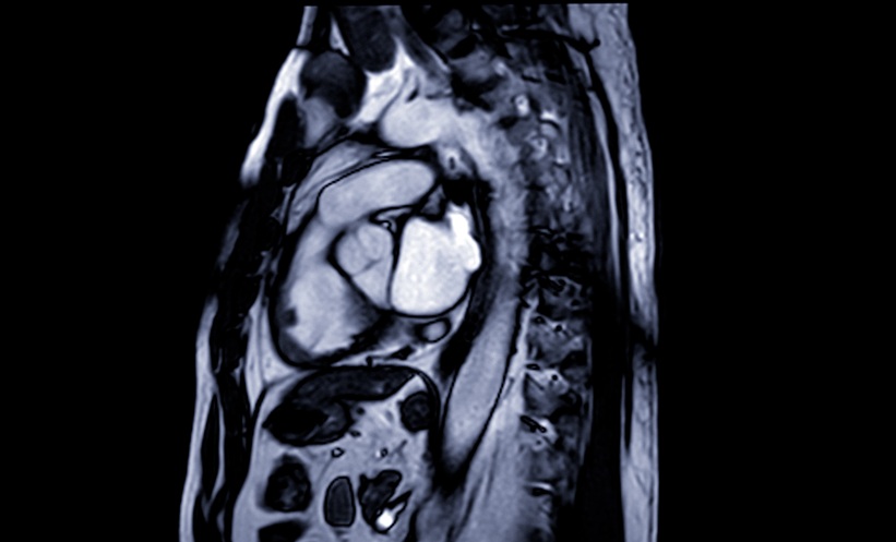
Researchers trained a machine learning approach to determine brain age based on T1-weighted MRI scans. The study used two datasets, from the Beijing Aging Brain Rejuvenation Initiative (BABRI) and the Alzheimer’s Disease Neuroimaging Initiative (ADNI). In total, 974 healthy controls were used for model training, with an age range of 49–95 years. Following this training, the model was tested on 231 healthy controls, and 224 patients with aMCI to estimate predicted age difference (PAD); its associations with cognitive impairment, genetic risk factors, and pathological markers of Alzheimer’s disease; and clinical progression. These factors were respectively analysed using a partial correlation analysis, a two-way analysis of covariance, and a general linear model.
Results from the prediction model showed that patients with aMCI were found to have higher PADs than those of healthy controls. PADs were significantly associated with individual cognitive impairment in multiple cognitive domain in the aMCI cohort. In terms of risk factors, apolipoprotein E ε4 allele carriers had higher PADs than non-carriers, and similarly, those with amyloid-positive aMCI had higher PADs than patients who were amyloid-negative. The use of PAD alongside other markers of Alzheimer’s disease aided the initial differentiation between progressive and stable aMCI, and this was reflected in an area under the curve value of 0.87.
The researchers concluded that PAD can be considered a sensitive imaging marker that is related to individual cognitive differences in patients with aMCI.