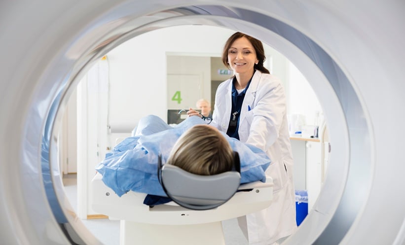
ORAL squamous cell carcinoma (OSCC) can invade the mandible, influencing the extent of surgery required. A retrospective study found that combining computed tomography (CT) and magnetic resonance imaging (MRI) increased sensitivity for detecting mandibular invasion, although overall diagnostic accuracy remained unchanged compared with either modality alone.
Comparing CT and MRI Accuracy
Researchers retrospectively analysed 57 patients with treatment-naïve OSCC whose tumours abutted the mandible. All patients underwent preoperative CT and MRI before surgical treatment between 2016 and 2021. Imaging findings were compared with histopathological examination of the resected mandible, which served as the reference standard.
Mandibular invasion was confirmed in 25 patients. CT demonstrated a sensitivity of 88.0% and specificity of 90.6%, while MRI achieved 92.0% sensitivity and 87.5% specificity. Both modalities had an overall diagnostic accuracy of 89.5%.
When either scan indicated invasion, combined imaging achieved 96.0% sensitivity and 96.4% negative predictive value. However, specificity decreased to 84.4%.
Implications for Surgical Planning
Researchers also assessed the detection of cortical and medullary involvement. Combined imaging identified cortical invasion with 88.0% sensitivity and 100% specificity, while sensitivity for medullary invasion was 92.0%, with specificity of 87.5%.
Researchers suggested that the higher negative predictive value of combined imaging could help identify patients who may not require more extensive resection. However, the study did not assess whether combining the modalities changed surgical decisions or patient outcomes.
Medullary Invasion Associated with Poorer Survival
Survival analysis showed that five-year overall survival was 87.4% among patients without mandibular invasion, compared with 55.5% in those with cortical invasion and 37.8% in those with medullary involvement.
The difference was statistically significant, but the analysis was unadjusted and did not account for potential confounding factors such as tumour size, nodal status or adjuvant treatment.
Researchers acknowledged that the retrospective design, small sample and lack of blinding between radiologists and pathologists limited the findings.
They concluded that CT and MRI may have complementary roles in preoperative assessment, while larger cohorts and prospective studies are needed to validate diagnostic performance and establish whether combined imaging improves surgical decision-making and patient outcomes.
Reference:
Rehal S et al. Diagnostic performance of CT and MRI, individually and in combination, in the assessment of mandibular invasion using histopathology as a reference standard. Cancers.2026;18(22):3649.