MATERNAL valacyclovir treatment was linked to fewer prenatal brain abnormalities in fetuses with congenital cytomegalovirus (cCMV) infection, according to a retrospective cohort study. Treated fetuses were more likely to have persistently normal MRI findings and showed no early minor brain lesions, supporting a potential protective effect on fetal brain involvement during pregnancy
cCMV is the leading cause of non-genetic sensorineural hearing loss and neurodevelopmental impairment worldwide. The infection affects an estimated 0.5% to 2% of live births, although prevalence varies geographically. While most affected infants are asymptomatic at birth, up to 15% may later develop long-term complications.
MRI Findings Highlight Potential Benefit
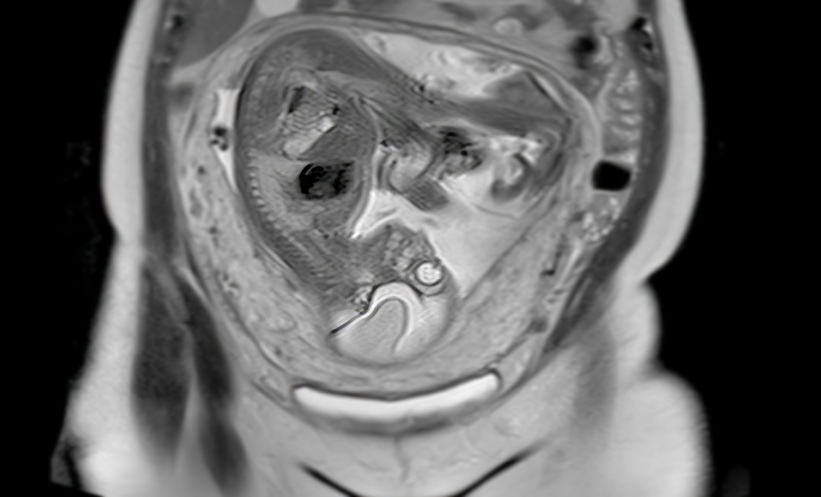
Prenatal MRI has become an important tool for the assessment of cCMV infection in utero, offering a greater sensitivity than ultrasound for detecting subtle brain abnormalities. These so-called minor lesions, including temporal pole T2 hyperintensity, focal temporal horn dilatation and small cysts, have been linked to later hearing impairment and mild neurodevelopmental difficulties.
Oral valacyclovir, administered to the mother at a dose of 8 g daily from diagnosis until delivery, is capable of achieving higher maternal and fetal drug concentrations than acyclovir and has been investigated as an antiviral strategy during pregnancy.
Fewer Early Brain Changes Observed
The single-centre study included 77 fetuses with confirmed cCMV infection who underwent at least two prenatal brain MRI examinations. Fetuses with major brain malformations at baseline were excluded.
Among the 15 treated fetuses, 73.3% had persistently negative MRI findings on serial imaging, compared with 37.1% of the 62 untreated fetuses. This difference was statistically significant (p=0.018; OR 4.67, 95% CI 1.29-16.9).
Notably, early-onset minor lesions, defined as abnormalities already visible on the first MRI examination, were only observed in untreated fetuses, affecting 11.3% of that group. When the analysis focused specifically on temporal pole lesions, the difference between treated and untreated fetuses remained significant (p=0.02).
What The Findings Could Mean
Lesion onset was categorised as absent, late or early, based on serial MRI assessments reviewed in consensus by two senior neuroradiologists. According to the investigators, maternal valacyclovir therapy was linked to a greater proportion of fetuses remaining MRI-negative throughout pregnancy and to the absence of early minor brain lesions.
Although the number of treated cases was relatively small, the statistically significant findings suggest a potentially meaningful signal that maternal antiviral therapy may help limit fetal brain involvement in cCMV. Further studies will be needed to confirm these observations and clarify their impact on longer-term neurological and hearing outcomes.
Reference
Tortora M et al. Serial prenatal MRI evaluation of valacyclovir-treated vs untreated fetuses with congenital cytomegalovirus (cCMV) infection. Eur Radiol. 2026;DOI:10.1007/s00330-026-12698-y.
Featured Image: Samunella on Adobe Stock
- Author: