BACKGROUND AND AIMS
Assessment of endometrial receptivity remains one of the major challenges in reproductive medicine despite significant advances in embryo selection and assisted reproductive technologies. At the 2026 European Society of Human Reproduction and Embryology (ESHRE) Annual Meeting, the authors presented a feasibility study evaluating serial intrauterine dissolved oxygen measurements throughout the luteal phase in healthy women.¹
The rationale for the study stems from experimental evidence suggesting that uterine oxygen tension varies dynamically during the peri-implantation period and may reflect underlying endometrial vascular remodelling.2-4 While previous human studies have reported isolated intrauterine oxygen measurements, longitudinal profiling across the entire luteal phase had not previously been described. Furthermore, embryonic metabolic requirements support a physiological role for dynamic intrauterine oxygen regulation. Early embryos rely predominantly on anaerobic metabolism during cleavage stages, whereas oxygen-dependent metabolism increases substantially at the blastocyst stage. Consequently, changes in intrauterine oxygen availability during the luteal phase may align with the changing metabolic demands of the developing embryo.⁵
MATERIALS AND METHODS
In this prospective observational pilot study, eight healthy women aged 18–35 years with regular menstrual cycles underwent serial intrauterine dissolved oxygen (pO₂) measurements during a natural menstrual cycle. Measurements were performed from luteinising hormone (LH) surge LH+1 to LH+13/14 using a fibre-optic microsensor positioned approximately 1 cm from the uterine fundus under ultrasound guidance. The primary objective was to evaluate the feasibility of repeated intrauterine oxygen assessment and to characterise temporal oxygenation patterns across the luteal phase.
RESULTS
The investigators successfully obtained serial measurements in all participants and identified two distinct oxygenation profiles. Four participants demonstrated a characteristic ‘peak’ pattern, with low oxygen tension during the early luteal phase followed by a marked increase between LH+4 and LH+6, reaching values of approximately 40–45 Torr before declining after LH+8. One of these participants exhibited a similar but temporally advanced peak. In contrast, four participants maintained relatively low oxygen levels throughout the luteal phase without a discernible mid-luteal rise, constituting a ‘no-peak’ pattern. The two distinct oxygenation patterns observed across the luteal phase are illustrated in Figure 1.
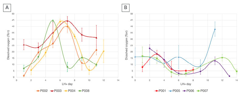
Figure 1: Representative intrauterine dissolved oxygen profiles during the human luteal phase.
Temporal profiles of intrauterine dissolved oxygen concentration (pO₂, Torr) across the luteal phase. The x-axis
represents days relative to the luteinising hormone surge (LH+ days), ranging from LH+0 to LH+14, and the y-axis shows intrauterine oxygen levels (0–50 Torr). Each coloured line represents an individual participant, with mean pO₂ values and error bars indicating standard deviations at each time point. pO₂ values represent averages obtained from a 5-minute continuous recording period at each time point. Participants are categorised into two distinct
response patterns:
A) Peak pattern: characterised by a mid-luteal rise in O₂ concentration (participants P002, P003, P004, and P008).
B) No-peak pattern: characterised by consistently low or stable pO₂ levels throughout the luteal phase (participants P001, P005, P006, and P007).
LH: luteinising hormone.
Post-hoc review identified potential physiological, pharmacological, or lifestyle-related factors among participants exhibiting the no-peak profile. Although causal relationships cannot be established from this small pilot cohort, these observations raise the possibility that intrauterine oxygen dynamics may be sensitive to factors influencing endometrial maturation and function.
Importantly, the procedure was well tolerated, with reported pain scores ranging from 0–2 on a visual analogue scale and no procedure-related adverse events. These findings support the technical feasibility and acceptability of serial intrauterine oxygen monitoring.
CONCLUSION
The study introduces a novel concept: the use of real-time intrauterine oxygen profiling as a functional biomarker of endometrial status. Unlike molecular or histological approaches, oxygen assessment provides an immediate physiological measurement obtained within the same menstrual cycle. If validated in larger studies and infertility populations, this approach could offer a minimally invasive method for evaluating embryo–endometrium synchrony and potentially refining embryo transfer timing.
Although limited by its small sample size and exploratory design, this work represents the first in vivo characterisation of dynamic intrauterine oxygen changes across the luteal phase in women and provides a foundation for future studies evaluating intrauterine oxygen profiling as a potential same-cycle functional biomarker of embryo–endometrium synchrony and endometrial receptivity.