Abstract
Background: Preterm birth is the main cause of neonatal morbidity and mortality, and its pathophysiology is only beginning to be understood. While it is considered a complex phenomenon where environmental and biological factors converge, inflammation has been identified as an important mechanism. Moreover, preterm birth has been classified as late preterm or early preterm, according to whether it occurs between Weeks 34–<37 or prior to Week 34, respectively. Early preterm carries significantly more risk for the newborn. Nonetheless, there are no known markers that help to make a differential prognosis, or to target in order to optimise prophylactic therapy with a precision medicine focus.
Methods: The authors analysed the cervicovaginal immune secretion patterns of a cohort of 133 pregnant individuals at high risk for preterm birth, and compared those who presented with term birth against a group with early preterm or late preterm births. The samples were taken at 20–24 weeks of pregnancy.
Results: The authors found that early preterm births are associated with reduced levels of IL-10 and granulocyte-macrophage colony-stimulating factor (GM-CSF), and enhanced levels of IL-8. On the other hand, late preterm birth was associated with increased IL-8, IL-6, IL-1β, C3b/iC3b, C5, IgE, and IgM. All markers except IL-10 and C5 displayed a good ability to work as predictors of their respective preterm birth category. No marker was found to be associated with vaginal dysbiosis.
Conclusion: Both early preterm birth and late preterm birth correlate to distinctive inflammatory patterns, which may show that both entities have different pathophysiological mechanisms.
Key Points
1. Babies that are born closer to their estimated time of delivery have the least risk for morbidity and mortality, but there is a lack of knowledge that allows prognosis and prevention.2. The present cohort study delves into the inflammatory and microbial factors associated with early or late preterm birth.
3. The authors show that different inflammatory patterns are associated with early and late preterm birth, pinpointing different pathophysiological mechanisms that may regulate such entities.
BACKGROUND
Spontaneous preterm birth (PTB) is a major cause of neonatal morbidity and mortality worldwide.1 PTB is defined as birth that occurs before 37 completed weeks of gestation. Approximately 13 million preterm babies are born each year, of whom about one million die before the age of 5 years; 35% of these deaths occur in infants younger than 28 days, since they have a higher risk of respiratory, infectious, and neurological complications, as well as long-term sequelae that significantly affect quality of life and health systems. They may present complications such as thermal instability, respiratory distress, infections, apnoea, hypoglycaemia, seizure episodes, jaundice, necrotising enterocolitis, periventricular leukomalacia, prolonged hospitalisation, and hospital readmission.2
Several risk factors for PTB have been determined, and the commonly recognised aetiologies include individual-level psychosocial and behavioural factors, neighbourhood characteristics, exposure to the environment, medical conditions and comorbidities, infertility treatments, biological factors, and genetics, but PTB is increasingly recognised as a biologically heterogeneous syndrome, in which inflammatory and host-microbiome interactions appear to play a major role.3-5 In fact, recent research has highlighted the role of certain receptors, chemokines, and inflammatory cytokines in cervical ripening, membrane rupture, and uterine contractility.6
Moreover, a gradient in the risk for adverse outcomes in the neonate has been observed, where those born closer to their estimated time of delivery have the least risk, while those born earlier have an enhanced risk. In such scenarios, PTB has been classified into early PTB for those births that occur before 34 weeks of gestation, and late PTB for those cases where the baby is born between Weeks 35–36.7-9 Despite early PTB being more prone to complications, little research has been done to understand its pathophysiology, to develop strategies for its timely recognition and prevention, and for its treatment.10
Evidence suggests that host-microbiome interactions and cervicovaginal inflammation, including complement activation as well as Ig production, contribute to the pathophysiology of spontaneous preterm birth.1 Nonetheless, the interactions between the aforementioned variables, and their roles and timing, are fields that are just being developed.11,12 Furthermore, fewer analyses have translated these findings into clinically relevant phenotypes such as early PTB and late PTB.12 Although some advancements in the detection of nearing births have been made,13,14 these tests are sensitive only 7 days after a patient starts to develop signs and symptoms of early labour.6 Thus, we currently lack the understanding to develop a test to accurately predict preterm birth with significant advancement and to discriminate the cases where an early or late PTB may occur.
On the other hand, precision medicine has delivered enhanced therapeutics and diagnostics in other fields, as it focuses on stratification of risk factors and pathophysiological mechanisms to ‘personalise’ medicine.15-17 In such a context, understanding the inflammatory mechanisms driving either early or late preterm deliveries may allow for better risk stratification and more precise therapeutic tools.
AIMS
The authors hypothesised that the inflammatory mechanisms regulating early and late preterm birth should be different. Thus, they aimed to describe the fundamental differences in the levels of inflammatory markers between individuals presenting with term births, early PTB, and late PTB. They then aimed to study the ability of each marker to work as a diagnostic/prognostic tool for the identification of preterm birth and studied the correlation of the significant markers with vaginal dysbiosis to understand more about the possible aetiologies of both early and/or late PTB.
METHODS
Study Design
A secondary analysis of an existing dataset derived from the March of Dimes (MOD) Database for Preterm Birth Research was performed (data descriptor available from Sirota et al.18). The MOD arose from prospectively recruited patients at five preterm birth prevention clinics in the UK.12
Participants
The cohort included 133 individuals with a high risk of PTB. Inclusion criteria were history of previous spontaneous PTB, midtrimester loss, and recurrent miscarriage, as well as the incidental finding of cervical shortening and/or cervical excisional treatment. Exclusion criteria were individuals under the age of 18 years, HIV or hepatitis C positive status, and vaginal intercourse or bleeding within 72 h of sample collection. Participants with iatrogenic preterm birth were eliminated.
Setting
Longitudinal cervicovaginal sample swabs were collected at timepoint A (12–16 weeks), timepoint B (20–24 weeks), and timepoint C (30–34 weeks). Luminex immunoassays (DiaSorin, Saluggia [Vercelli], Italy) were performed to determine the levels of IL-1β, IL-2, IL-4, IL-5, IL-6, IL-10, IL-18, interferon (IFN)-γ, granulocyte-macrophage colony-stimulating factor (GM-CSF), and TNF-α for cytokines; IgM, IgG1, IgG2, IgG3, IgG4, IgA, and IgE for Igs; and C3b/iC3b, C5, and C5a for complement proteins. 16S ribosomal RNA sequencing was performed to determine the microbiological profile of each individual, and individuals were classified according to the Valencia criteria for vaginal microbiota. All methods were carried out as described in Chan D et al.12
For this analysis, PTB was defined as delivery before 37+0 weeks, early PTB as delivery before 34+0 weeks, and late PTB as delivery between 34+0 and 36+6 weeks, consistent with CDC and American College of Obstetricians and Gynecologists (ACOG) definitions.4
Data Collection
The authors of the original study deposited the data at the ImmPort database and data-sharing portal.19 For the current study, these data were downloaded from ImmPort.
Data Analysis
Comparisons of immune mediator levels were performed across term birth, early PTB, and late PTB groups. A Shapiro–Wilk test was performed to assess for normality, and data with p≤0.05 were considered non-parametric. The authors next performed a Mann–Whitney U test to compare the levels of Igs, cytokines, and complement proteins among the individuals reaching term birth, late PTB, or early PTB.
A Chi-square test was used to assess the association between vaginal dysbiosis and PTB, and odds ratios with CI were calculated, considering p≤0.05 and CI that did not include the value of 1 as significant.
Spearman correlation analysis wasused to explore associations betweeninflammatory mediators and Lactobacillusspp. abundance. Correlation coefficients were classified as strong (r≥0.7), good or moderate (r=0.5–0.7), weak (r=0.3–0.5), or very weak (r≤0.3).20
To evaluate predictive performance, receiving operator characteristics (ROC) curve analyses were performed for selected biomarkers measured at period B, and area under the ROC (AUROC) values were calculated, considering markers as good predictors when p≤0.05.
All data analysis and plotting were performed using GraphPad Prism X9 (GraphPad Software, San Diego, California, USA).
Ethical Approval
The parent study received approval from the appropriate Institutional Review Board, and all participants provided written informed consent before enrolment. This secondary analysis was conducted using data derived from that approved cohort. The secondary analysis was approved by the Comité de Ética en Investigación del Instituto de Ciencias de la Salud (CEI-ICSa), with the approval number CEI-ICSa-2025/R007.
FINDINGS
No significant difference in maternal age (18–46 years [29.7±6]; p=0.93), BMI (p=0.20), or ethnicity (p=0.09) were found among individuals presenting with term birth or PTB.18 The analysis of the secretion patterns of cytokines (Figure 1), complement proteins (Figure 2), and Igs (Figure 3) revealed distinctive patterns for both early and late PTB. While early PTB was associated with a decreased expression of IL-10 (Figure 1A) and GM-CSF (Figure 1B), and an enhanced expression of IL-8 (Figure 1C), late PTB was found to be associated with enhanced levels of IL-8 (Figure 1C), IL-6 (Figure 1D), IL-1β (Figure 1E), C3b/iC3b (Figure 1F), C5 (Figure 1G), IgE (Figure 1H), and IgM (Figure 1I). The authors did not find significant differences between IL-2, IL-5, TNF-α, IL-18, C5a, IgA, IgG1, IgG2, IgG3, or IgG4 (data not shown).
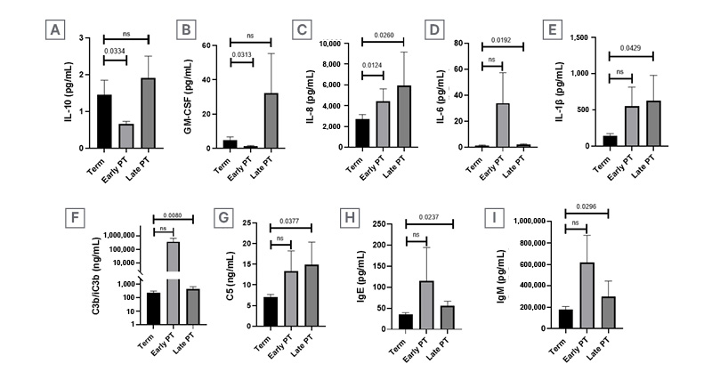
Figure 1: Cervicovaginal inflammatory secretion patterns are different for either early or late PTB.
The levels of cervicovaginal IL-10 (A), GM-CSF (B), IL-8 (C), IL-6 (D), IL-1β (E), C3b/iC3b (F), C5 (G), and
cervicovaginal IgE (H) and IgM (I), were measured and compared between individuals who gave birth either at term, early PT, or late PT.
GM-CSF: granulocyte-monocyte colony-stimulating factor; NS: not significant; PT: preterm; PTB: preterm birth.
Furthermore, a χ² test showed no correlation between vaginal dysbiosis and preterm birth in the author’s cohort (odds ratio: 1.41; CI: 0.47–4.20; p=0.52), but by means of a Spearman correlation the authors found a weak but significant negative correlation of IL-1β with Lactobacillus crispatus (r=0.36; p=0.00002; Figure 2).
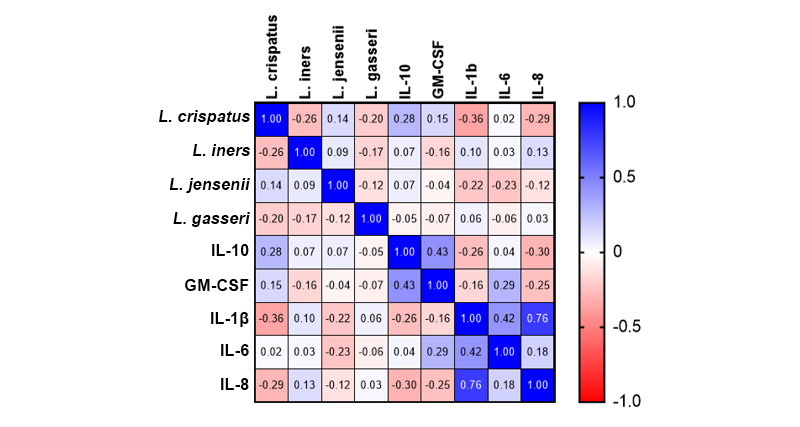
Figure 2: Cervicovaginal cytokines negatively correlate to Lactobacillus spp.
A Spearman multi-variable correlation was calculated for IL-1β, IL-6, IL-8, GM-CSF, and IL-10 with Lactobacillus crispatus, L. iners, L. jensenii, and L. gasseri.
Moreover, when the values of cytokines, Igs, and complement proteins of individuals presenting PTB (both in early or late stages) were compared to those of individuals that presented term birth (Figure 3), the authors found that GM-CSF (Figure 3B), IL-1β (Figure 3C), IL-8 (Figure 3D), IL-6 (Figure 3E), C3b/iC3B (Figure 3F), IgE (Figure 3H), and IgM (Figure 3I) had significant prognostic values, as calculated by a ROC curve test. Only IL-10 (Figure 3A) and C5 (Figure 3G) did not exhibit significant AUROC values.
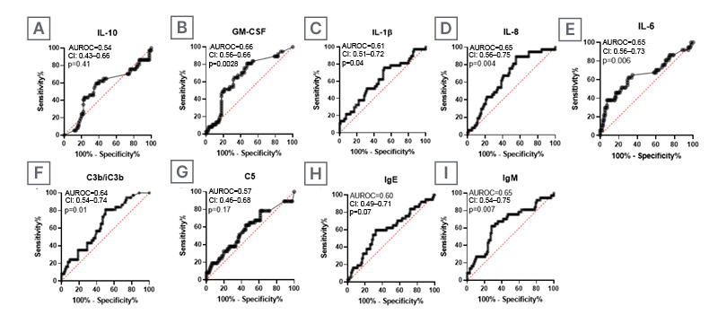
Figure 3: Most inflammatory markers exhibit significant sensitivity to predict preterm birth.
The levels at period B of cervicovaginal IL-10 (A), GM-CSF (B), IL-1β (C), IL-8 (D), IL-6 (E), C3b/iC3B (F), C5 (G), IgE (H), and IgM (I) were compared in individuals with preterm birth at both early and late stages to individuals with term birth to calculate the sensitivity of each marker.
GM-CSF: granulocyte-monocyte colony-stimulating factor; AUROC: area under the receiver operating characteristic curve.
DISCUSSION
The present research shows that different underlying immunological mechanisms associate to either early or late PTB. The mechanism behind early PTBs appears to be dependent on decreased IL-10 and GM-CSF production, as well as enhanced IL-8 production. On the other hand, IL-6, IL-8, IL-1β, C3b/iC3b, C5, IgE, and IgM are significantly enhanced in individuals who delivered at late preterm. Moreover, all these markers, except IL-10 and C5, exhibit significant sensitivity to act as predictors for PTB.
Although PTB has been described as primarily being caused by infections, multiple gestations (twins/triplets), placental complications, or chronic maternal health conditions, among others, the exact cause of more than 50% of preterm deliveries is never precisely determined.5 In such a setting, inflammation has appeared as an important factor that may explain many cases, and its role in the regulation of birth timing is only beginning to be understood.6,21
For instance, the fetus is a semi-allogeneic product; thus, pregnancy must induce a paradoxical immunological state where immune tolerance to the fetus co-exists with immune surveillance and defence. IL-10 has been linked to the induction of such immunotolerance, while the loss of IL-10-producing cells has been demonstrated to correlate with fetal rejection.22-24 In such a scenario, reduced IL-10 production is expected to induce product rejection and thus early PTB.
Nonetheless, GM-CSF is known to accelerate membrane rupture as well as cervical remodelling, thus precipitating deliveries.25,26 Therefore, its lower levels in individuals with early PTB are a finding that requires further investigation. On the other hand, cytokines like IL-1β and IL-6, and chemokines like IL-8, have been shown to play important roles in the preterm induction of birth.27 However, the authors did not find enhanced IL-1β or IL-6 levels in individuals with early PTB, and the only pro-inflammatory marker that was over-expressed in such settings was IL-8. In such circumstances, the authors think that increased inflammation in concordance with a loss of tolerance may induce early fetal rejection.
Moreover, the authors found that the local immunological profile of individuals with late PTB consists of high levels of IL-6, IL-8, and IL-1β, as well as C3b/iC3b, C5, IgE, and IgM. Such a profile is consistent with an infection-associated acute Th1 immune response at a mucosal interface, but the authors mostly failed to trace its origin to vaginal dysbiosis, only finding a significant negative correlation of IL-1β with L. crispatus.28,29 Such an experiment was not able to confirm that IL-1β correlates with dysbiosis, because it did not seem to be negatively correlated to the other Lactobacillus species. However, most markers exhibited an enhanced ability to work as markers for prediction of PTBs.
Interestingly, a shift from a tolerogenic Th2-type immune response to a Th1-type inflammation has been described as a phenomenon that occurs prior to delivery, and that is thought to induce changes at the maternal–fetal interface to induce labour.30,31 It has been shown that this shift, whenever it occurs during pregnancy, is able to induce labour, thus associating with PTBs and even miscarriage.32-34
Recent and pioneering research by Bezirganoglu-Altuntas et al.35 found that, while neutrophil-to-lymphocyte ratio and systemic immune inflammation index are good predictors for PTBs, there is no specific marker to distinguish between early and late PTBs. And, to the authors’ knowledge, no other research efforts have been made in order to discover differences in the pathophysiology of early versus late PTB. In such an instance, this is, to their knowledge, the first report about distinctive traits for early versus late PTBs.
IMPLICATIONS FOR PRACTICE
Although the role of inflammation, especially of IL-1β, IL-6, IL-8, TNF-α,36,37 C3b/iC3b, C5, and IgM, in the pathophysiology of PTB has been established,12 we currently lack much detail about the interplay between such markers and the specific timing for delivery. In such a context, the authors are unable to stratify the risk for PTB, and thus cannot calculate the urgency for interventions.
Precision medicine is considered a powerful strategy to enhance treatment success, as it is derived from the integration of big data, AI, various omics, and the study of environmental and social factors, and its objective is to tailor treatments to subpopulations that present a common trait.38 In such an understanding, the discrimination of the mechanisms that may induce PTB either at early or late stages represents a valuable tool for precision medicine. Such distinction could enhance the efficacy of therapies that depend on the modulation of the aforementioned inflammatory markers, such as IL-6.39 Moreover, efforts to predict intra-amniotic infection and inflammation with non-invasive tests may benefit from the knowledge that not all the inflammatory mechanisms that drive preterm delivery work in the same way, or at the same timepoints during pregnancy.40
Thus, the characterisation of distinctive inflammatory profiles for either early or late preterm deliveries may serve to 1) develop precise prognostic tests to assess and stratify the risk, and 2) develop tailored treatments with an enhanced ability to abrogate the pathophysiological mechanisms behind each type of preterm delivery, and thus be more effective at PTB prevention.
LIMITATIONS
Although the authors measured the correlation between the inflammatory status and the vaginal microbiota, the present study is limited by the lack of information regarding the chorioamnionitis status. This hinders the potential for the interpretation of the findings. Moreover, all the individuals in the cohort were at risk of PTB, so the control group does not represent the baseline characteristics of a normal pregnancy. Finally, even when the sample was sufficient to find significant differences, the authors cannot assure that the sample is of adequate size; thus, the results shown here should be interpreted with care.
CONCLUSION
The inflammatory patterns for individuals presenting early PTB are radically different from those presenting late preterm deliveries. The first consists not only of enhanced inflammation, but also has elements suggestive of a loss of tolerance. On the other hand, the latter is characterised only by frank inflammation.