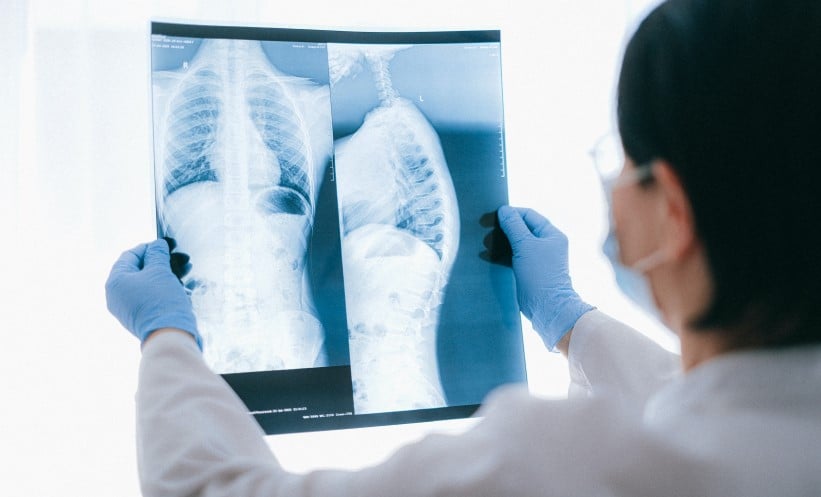
RESEARCHERS in France have discovered that typical chest CT scan patterns seen in patients with COVID-19 appeared much less frequently in individuals who had an Omicron infection, or were vaccinated. The retrospective multicentre study also showed that patients who had received more vaccine doses tended to display less severe results on CTs.
A cohort of almost 4,000 patients who had confirmed severe respiratory syndrome coronavirus 2, and had CTs taken of their chests, were analysed. A multivariable analysis pinpointed that patients who were infected with the Omicron strain of COVID-19 were 54% less likely to have typical CT findings associated with COVID-19, versus those with Delta infection (odds ratio [OR]: 0.46; 95% confidence interval [CI]: 0.37–0.57).
In typical CT scans of patients with COVID-19, practitioners expect to see the following: consolidations, fibrotic bands, intralobular reticulation, and ground glass opacities. All of these were less frequent in patients who had received two or three vaccine doses (OR: 0.32; 95% CI: 0.25–0.41, and OR: 0.20; 95% CI: 0.15–0.27, respectively).
Lead study author Guillaume Gorincour, Centre Hospitalier Universitaire Édouard Herriot, Lyon, France, and team also found that high severity scores, as in accordance with the French Society of Radiology-Society of Thoracic Imaging (SFR-SIT), were between 53–67% less likely in patients who had previously been given two or three doses of vaccine (OR: 0.47; 95% CI: 0.36–0.60, and OR: 0.33; 95% CI: 0.24–0.46, respectively). These results were irrespective of severe respiratory syndrome coronavirus 2 variant, sex, age, or existing comorbidities.
Gorincour stated: “Our results confirm that both the Omicron variant and vaccination were associated with lower frequency of typical chest CT for COVID-19 and lesser extent of disease, with a higher number of vaccine doses showing an increasing protective impact against high severity score.”
Limitations of the study are the collection of data from emergency departments, which may not be entirely accurate due to high workload. Reports were based on the SFR-SIT guidelines, and thus bronchial wall thickening and peribronchovascular predilection were not evaluated, despite being more frequent markers of the Omicron variant. There is also the potential for bias amongst radiologists, as awareness of their patient’s vaccination status may have skewed their interpretation. Chest CTs were also not routinely carried out in COVID-19 cases.


