BACKGROUND AND AIMS
Severe constitutional genu varum in young adults often leads to abnormal mechanical loading across the knee, accelerating medial compartment osteoarthritis. The primary goal of surgical treatment is to restore a near-normal mechanical axis and delay joint degeneration. This report evaluates the rationale, technique, and outcomes of simultaneous double femoral and tibial osteotomy performed in a single operative session for severe varus malalignment.1
METHODS
The authors present the case of a 30-year-old male with dwarfism secondary to multiple epiphyseal dysplasia, presenting with bilateral knee pain and an intercondylar distance of 25 cm. Clinical examination showed preserved range of motion. Radiological assessment revealed preserved joint spaces but severe varus deformity, with a hip-knee-ankle (HKA) angle of 149° on the right and 145° on the left. A simultaneous double osteotomy was performed in one operative session per limb. The procedure consisted of a lateral closing-wedge distal femoral valgus osteotomy, stabilised with a Dynamic Condylar Screw plate (DePuy Synthes, Warsaw, Indiana, USA),2 followed by a medial opening-wedge proximal tibial valgus osteotomy, stabilised with a T-plate.3 Importantly, the bone wedge resected from the femur was used as an autograft for the tibial opening wedge. Partial weight-bearing began on Day 45, with full weight-bearing permitted at 3 months after radiological confirmation of union. The contralateral limb was treated with the same technique 1 year later.
RESULTS
Postoperative recovery was uneventful, with no infections, non-unions, or neurovascular complications. At the 1-year follow-up, the patient reported high functional satisfaction with both knees, with significant improvement in gait and pain reduction. Although a slight residual varus was noted on standing long-leg radiographs, the overall mechanical axis was substantially corrected (Figure 1)). The use of the femoral autograft eliminated donor-site morbidity and facilitated tibial healing.
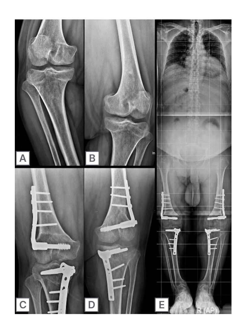
Figure 1: Pre-operative, postoperative, and long-term radiographic follow-up.
Significant pre-operative deformity with bilateral genu varum (A, B).
Simultaneous osteotomy performed in a single procedure: lateral femoral closing wedge osteotomy and medial tibial opening wedge osteotomy for the right knee (C), and the same procedure for the left knee 1 year later (D).
Telemetric radiograph at the latest follow-up shows good union at the osteotomy sites and satisfactory alignment of both lower limbs (E).
CONCLUSION
Combined distal femoral and proximal tibial osteotomy for severe genu varum is a complex but effective procedure. Performing both osteotomies simultaneously reduces the number of anaesthetic exposures, shortens overall treatment duration, and accelerates rehabilitation compared to staged surgeries. The technique allows for precise correction of deformity at its anatomical origins (femoral and tibial) rather than attempting compensatory correction at a single level, which can lead to joint line obliquity. Computer navigation, when available, improves precision, reproducibility, and real-time feedback during correction.
Simultaneous double femoral and tibial osteotomy is a highly effective option for young adults with severe genu varum, particularly when deformity arises from both bones. When it is well mastered and assisted by navigation, the procedure provides reliable realignment, delays osteoarthritis, and yields high patient satisfaction.