Abstract
Rosacea is a chronic, multifactorial inflammatory dermatosis involving innate immune dysregulation, neurovascular instability, and microbiome imbalance. Severe papulopustular phenotypes often relapse and exhibit resistance to conventional topical therapy, creating a need for antibiotic-sparing strategies aligned with antimicrobial stewardship.
The authors report a 45-year-old female with severe, recurrent papulopustular rosacea refractory to topical ivermectin and metronidazole. An integrative, antibiotic-free protocol was implemented, combining a functional dermocosmetic regimen (barrier repair, mineral photoprotection) with oral probiotics for microbiome modulation.
Complete remission of inflammatory lesions occurred by Week 6, with sustained clearance at 6-month follow-up. No adverse events were observed, and patient adherence was excellent.
This case highlights the potential of multimodal dermocosmetic maintenance combined with microbiome support as an effective alternative to systemic antibiotics in severe rosacea, supporting phenotype-driven care and antimicrobial stewardship principles.
Key Points
1.Rosacea affects approximately 5% of adults; papulopustular disease frequently relapses after topical therapy is stopped, and repeated courses of systemic antibiotics raise antimicrobial stewardship concerns.2. This article discusses the case of a 45-year-old woman with severe, recurrent papulopustular rosacea refractory to topical ivermectin and metronidazole, treated with an antibiotic-free protocol combining a multimodal dermocosmetic regimen (barrier repair, vascular stabilisation, mineral photoprotection) with oral probiotics targeting the gut–skin axis.
3. Complete remission of inflammatory lesions was achieved by Week 6 and was sustained at 6-month follow-up without systemic antibiotics, suggesting that barrier- and microbiome-focused maintenance may be a valid antibiotic-sparing option in selected patients; controlled trials are needed to confirm efficacy.
INTRODUCTION
Rosacea is a chronic inflammatory dermatosis affecting approximately 5% of the adult population, manifesting through erythematotelangiectatic, papulopustular, phymatous, and ocular phenotypes.1,2 As a chronic, relapsing–remitting condition, rosacea currently has no established cure; therapeutic goals are centred on achieving and maintaining control of signs and symptoms, since remission can be interrupted by relapse even after successful treatment.1,3 First-line therapeutic strategies typically include topical metronidazole, ivermectin, and azelaic acid.4,5 In moderate-to-severe presentations, modified-release doxycycline (an anti-inflammatory, sub-antimicrobial dose of 30–40 mg immediate-release plus 10 mg delayed-release) is frequently employed, while light- and laser-based devices are reserved for persistent telangiectasia.5,6 At this sub-antimicrobial dose, plasma concentrations remain below the minimum inhibitory concentration for common pathogens, so the drug does not exert the selective pressure that favours resistant bacterial strains; long-term studies of continuous use have found no emergence of antibiotic-resistant organisms in oral, intestinal, or cutaneous flora with this regimen.5,7
However, disease recurrence upon treatment cessation is common, underscoring the need for effective maintenance strategies and adjunctive care, particularly gentle cleansing, barrier reinforcement, and rigorous photoprotection.
The pathogenesis of rosacea is multifactorial, involving complex, interconnected pathways (Figure 1).1,8,9 The first pathway, innate immunity, involves activation of toll-like receptor 2 (TLR2), a pattern-recognition receptor of innate immunity, in keratinocytes and mast cells.8-10 This leads to increased expression of kallikrein 5 (KLK5), a serine protease involved in cathelicidin processing, and subsequent cleavage of cathelicidin into LL-37, an antimicrobial peptide that promotes inflammation.8,9,11 LL-37 drives chemotaxis, angiogenesis, and inflammation via NF-κB, a pro-inflammatory transcription factor, and MAPK, a signalling pathway regulating inflammation.8,9
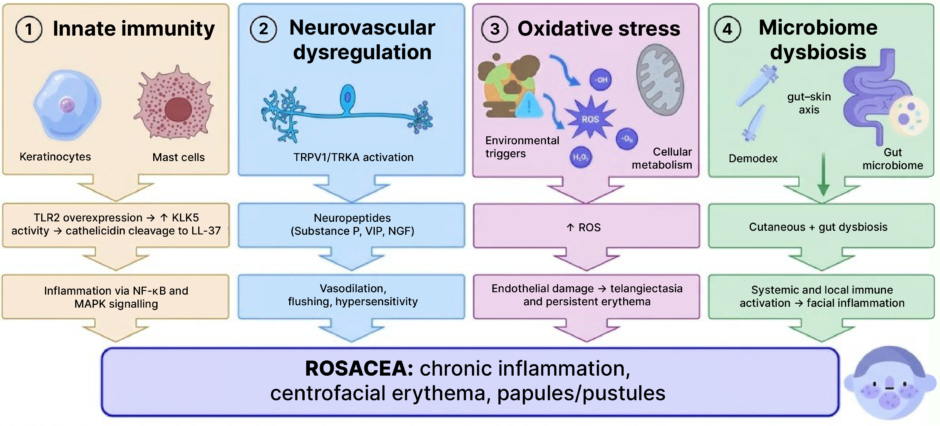
Figure 1: Rosacea pathogenesis: interconnected mechanisms driving chronic inflammation.
The four interconnected pathogenic pathways described in the introduction converge on the clinical phenotype of chronic inflammation, centrofacial erythema, and papules/pustules. KLK5: kallikrein 5; NGF: nerve growth factor; ROS: reactive oxygen species; TLR2: toll-like receptor 2; TRKA: tropomyosin receptor kinase A; TRPV1: transient receptor potential vanilloid 1; VIP: vasoactive intestinal peptide.
The second pathway, neurovascular dysregulation, is mediated by transient receptor potential vanilloid 1 (TRPV1), tropomyosin receptor kinase A (TRKA), a receptor for nerve growth factor, and neuropeptides such as nerve growth factor (NGF), substance P (a neuropeptide involved in pain and inflammation), and vasoactive intestinal peptide (VIP), which amplifies vasodilation and hypersensitivity.8,12
The third pathway, oxidative stress, characterised by increased reactive oxygen species (ROS) molecules that cause endothelial damage, contributes to persistent erythema.9
The fourth pathway, microbiome dysbiosis, involves cutaneous dysbiosis (a proliferation of Demodex folliculorum and Bacillus oleronius, and an imbalance of Staphylococcus epidermidis and Cutibacterium acnes) and gut microbiome alterations, which contribute to systemic inflammation via the gut–skin axis.2,13-16
These pathophysiological cascades explain the chronicity of the disease and support the rationale for multimodal therapeutic approaches.
CLINICAL CASE
A 45-year-old woman with no relevant past medical history presented with long-standing rosacea characterised by frequent relapses. On examination, there was diffuse, confluent erythema involving the malar region and lateral nasal sidewall, with irregular borders and a clear transition towards relatively unaffected mandibular and perioral skin, demonstrating a characteristic centrofacial distribution pattern consistent with persistent centrofacial vascular hyperreactivity. Prominent telangiectasias were noted, along with multiple inflammatory papules, clustered pustules with purulent exudate, and small plaques overlying an erythematous background with fine peripheral scaling. The lesions displayed variable chromatic intensity, ranging from light erythema to deep erythematous–violaceous discolouration, reflecting heterogeneous inflammatory activity. Areas of superficial scale, focal desquamation, and mild crust or micro-crust formation were evident, with scattered excoriations and superficial erosions, particularly in the perinasal region. The skin surface appeared irregular, thickened, and mildly oedematous in certain areas, with increased shine suggestive of epidermal barrier disruption. Cutaneous texture was notably altered, with visible and dilated follicular ostia, accentuated skin lines, and perifollicular inflammation. Fine vellus hairs and sebaceous skin characteristics were discernible under the lighting. Overall, the clinical appearance demonstrated prominent inflammatory, vascular, and epidermal alterations consistent with moderate-to-severe rosacea (Figure 2). The patient reported transient improvement with previous topical ivermectin and metronidazole therapy, followed by recurrent exacerbations after treatment discontinuation. She expressed a strong preference to avoid systemic antibiotics and requested an integrative, non-antibiotic therapeutic approach.
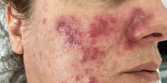
Figure 2: Papulopustular and erythematotelangiectatic rosacea.
Right hemiface at baseline, showing diffuse centrofacial erythema, clustered inflammatory papules and pustules with scale and crusting, and epidermal barrier disruption.
INTERVENTION
A multimodal dermocosmetic regimen was implemented to address key pathogenic mechanisms of rosacea, focusing on barrier repair, microbiome modulation, vascular stabilisation, and mineral photoprotection.17,18 The protocol included a cream–oil cleanser enriched with squalane (a skin-identical lipid that reduces transepidermal water loss), Lactobacillus ferment (a postbiotic for microbiome balance), and an antioxidant and anti-pollution complex to reinforce barrier integrity and mitigate oxidative stress. A soothing concentrate contained aloe vera, panthenol, acetyl tetrapeptide-2 (an antioxidant and anti-stress peptide), and acetyl heptapeptide-4 (providing microbiome support) to downregulate inflammatory mediators and restore barrier function. A vasoprotective moisturiser combined melilotus, ruscus, bilberry, hesperidin, and escin to reduce capillary permeability and inflammation, while squalane and panthenol restored hydration and lipid balance, targeting endothelial dysfunction and vascular fragility. Daily photoprotection was ensured with a mineral sunscreen with sun protection factor 50+ (SPF50+), formulated with zinc oxide and titanium dioxide (100% mineral filters), prebiotics (inulin and yoghurt powder), and botanical antioxidants to attenuate UV-induced oxidative stress and support microbiota balance. Finally, an oral probiotic supplement provided probiotics (Lactiplantibacillus plantarum, Limosilactobacillus reuteri, Lacticaseibacillus rhamnosus) combined with inulin, zinc, and biotin to modulate the gut–skin axis and immune signalling.2,13,14 In view of the relapse pattern and patient preference, systemic antibiotics were avoided, reserving modified-release doxycycline as a step-up option, but this was not required.5
OUTCOME
At the 6-week follow-up, there was complete resolution of the previously observed pustular lesions, and a marked reduction in diffuse centrofacial erythema, with significant attenuation of inflammatory activity in the malar and perinasal regions. Compared with the baseline presentation, the skin tone appeared more homogeneous, with substantial resolution of papules, plaques, and superficial crusting. Residual erythema was mild and localised, consistent with improved vascular stability and effective control of the inflammatory component of the disease. The epidermal surface showed restoration of barrier integrity, with diminished scaling, reduced textural irregularities, and decreased cutaneous oedema. Skin texture appeared smoother, with normalisation of follicular ostia visibility and reduced perifollicular inflammation. No pustules, erosions, excoriations, or other active inflammatory lesions were visible. By Week 12, clinical stabilisation was confirmed, with no evidence of new inflammatory activity and excellent tolerability of the therapeutic regimen (Figure 3). At 6 months, sustained remission was maintained, with persistent clearance of lesions and no infectious complications. Adherence to the prescribed dermocosmetic and probiotic protocol remained high throughout the observation period.
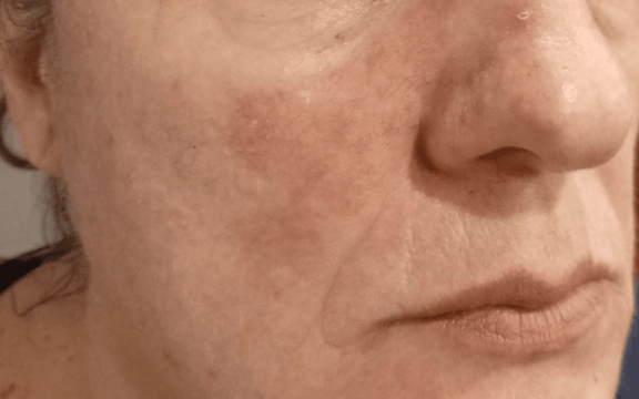
Figure 3: Clinical outcome after therapeutic protocol.
Right hemiface after completion of the therapeutic protocol, showing marked reduction of centrofacial erythema and resolution of papules and crusting, consistent with the sustained remission described in the outcome section above.
DISCUSSION
The sustained remission observed in this case can be attributed to the multimodal regimen targeting the four major pathogenic pathways of rosacea.
Innate Immunity and LL‑37 Axis
Overexpression of TLR2 and increased KLK5 activity lead to the excessive processing of cathelicidin into LL-37, promoting chemotaxis, angiogenesis, and inflammation via NF-κB and MAPK pathways.8–11 Barrier-supportive actives such as squalane (present in the cleanser and the vasoprotective moisturiser) restore lipid integrity and reduce transepidermal water loss, limiting irritant penetration and indirectly tempering TLR2 activation. Aloe vera and panthenol (in the soothing concentrate and vasoprotective moisturiser) exert anti-inflammatory and soothing effects, while acetyl tetrapeptide-2 provides antioxidant protection and stabilises the extracellular matrix, mitigating downstream inflammatory signalling.
Neurovascular Dysregulation
TRPV1/TRKA signalling and neuropeptides (NGF, substance P, VIP) amplify vasodilation and flushing.8,12 Vasoprotective phytocomplexes in the vasoprotective moisturiser (melilotus, ruscus, bilberry, hesperidin, and escin) strengthen capillary walls, reduce permeability, and improve microcirculation, counteracting neurovascular fragility. Panthenol further supports endothelial repair and hydration, reducing vascular reactivity.
Oxidative Stress
UV-driven ROS perpetuate erythema and telangiectasia. Strict mineral photoprotection with zinc oxide and titanium dioxide (from the mineral sunscreen) offers broad-spectrum UVA/UVB defence, preventing ROS generation.9 The antioxidant/anti-pollution complex in the cleanser neutralises pollution-related oxidative stress, while botanical antioxidants (helianthus, pongamia, lupinus, rosmarinus) in the mineral sunscreen scavenge free radicals and protect vascular endothelium. Acetyl tetrapeptide-2 in the soothing concentrate adds further antioxidant defence.
Microbiome Dysbiosis
Increased Demodex density and associated bacteria such as Bacillus oleronius correlate with disease exacerbations, while gut dysbiosis reinforces systemic inflammation along the gut–skin axis.14,15 Lactobacillus ferment (in the cleanser) acts as a postbiotic, promoting cutaneous microbiome homeostasis, and acetyl heptapeptide-4 (in the soothing concentrate) supports microbial balance on skin. Prebiotics such as inulin and yoghurt powder in the mineral sunscreen help maintain healthy microbiota under photoprotection. Oral supplementation with the probiotic supplement provides probiotics (Lactiplantibacillus plantarum, Limosilactobacillus reuteri, Lacticaseibacillus rhamnosus) and inulin to modulate gut microbiome and immune signalling, while zinc and biotin support immune regulation and skin integrity.2,13,14,16
By simultaneously targeting barrier dysfunction, oxidative stress, neurovascular instability, and microbiome imbalance, this regimen aligns with phenotype-driven management and antibiotic stewardship principles. Although evidence for probiotics and dermocosmetic adjuncts is growing, controlled trials are needed to validate efficacy and define optimal protocols.1,2,13,14,19
Clinical Implications
For patients who are unwilling or not suitable for systemic antibiotics, and/or those with frequent recurrence, structured maintenance centred on barrier care, vascular stabilisation, and photoprotection may provide durable control.1,19
The regimen described can be adapted with equivalent formulations; specific commercial product identities are disclosed separately, for transparency and reproducibility only, and do not imply endorsement.
Limitations
The limitations of this case report include: it being a single‑patient report without objective scoring; potential placebo and Hawthorne effects; the use of commercial dermocosmetics combining active ingredients in proprietary concentrations and associations limiting generalisability to other formulations with the same generic actives; and the absence of dermoscopy or microbiome assays. Controlled studies comparing barrier/microbiome‑focused maintenance versus standard care are warranted. Finally, rosacea is an inherently chronic, relapsing–remitting disease, and even complete clearance of lesions does not equate to a cure; relapse remains possible at any point, including after prolonged remission, and the sustained control achieved in this case should not be construed as a cure or as a guarantee of durable, antibiotic-free control in other patients.3
CONCLUSION
In this patient with severe papulopustular rosacea and recurrent pustular exacerbations, a structured maintenance strategy centred on barrier restoration, microbiome support, vascular stabilisation, and strict mineral photoprotection, supplemented by oral probiotics, achieved sustained remission at 6-month follow-up without the need for systemic antibiotics.2,13,14 This approach may not suit patients with frequent pustular recurrence and/or moderate-to-severe activity unresponsive to dermocosmetic and probiotic measures alone, for whom step-up doxycycline or other guideline-directed therapy remains indicated. These findings reinforce the value of phenotype-guided management and antibiotic stewardship, highlighting the potential role of multimodal dermocosmetic protocols in reducing relapses and improving long-term disease control in appropriately selected patients.