Abstract
Lupus erythematosus–lichen planus (LE–LP) overlap syndrome is a rare clinical disorder, which is characterised by the features of both LE and LP. The authors report a case of a 35-year-old male presenting with an itchy, verrucous annular plaque over the left elbow that had been present for 4 months. There was no history of any pain. Clinical diagnosis was challenging due to the atypical presentation, but histopathology revealed hyperkeratosis, wedge-shaped hypergranulosis, saw-tooth acanthosis, vacuolar degeneration, lymphocytic infiltrates, pigment incontinence, and mucin deposition in the dermis. Immunological evaluation showed positive antinuclear antibody and direct immunofluorescence with granular immunoglobulin and complement component 3 (C3) deposition and linear fibrinogen at the basement membrane zone. The patient was diagnosed with LE–LP overlap syndrome and treated with topical 0.5% clobetasol propionate ointment, and achieved rapid resolution within 3 weeks. This case emphasises the diagnostic complexity of LE–LP overlap syndrome, highlighting the need for histopathological and immunological correlation.
Key Points
1. This case highlights the importance of integrating all the clinical, histopathological, and immunological data to diagnose lupus erythematosus–lichen planus overlap syndrome.2. This is a case report presenting an unusual presentation on a non-sun-exposed site, emphasising the need for high clinical suspicion and comprehensive evaluation.
3. Clinicians should consider this entity in atypical verrucous plaques or annular lesions, even in non-sun-exposed areas, to avoid diagnostic delays.
INTRODUCTION
Lupus erythematosus (LE) and lichen planus (LP) are chronic immune-mediated inflammatory dermatoses. Although classically regarded as distinct entities, they share several clinical, histopathological, and immunopathogenic features.1 LE represents a disease spectrum ranging from cutaneous-limited variants like acute, subacute, and chronic cutaneous LE to systemic LE, a potentially life-threatening multisystem disorder.1,2 In contrast, LP is a T cell-mediated inflammatory disease, which primarily affects the skin and mucous membranes, and is characterised by pruritic, polygonal, violaceous papules and plaques with a predilection for flexural surfaces and mucosa.3
Despite their different clinical paradigms, both LE and LP are united by the presence of interface dermatitis, a histopathological reaction pattern defined by basal keratinocyte damage, vacuolar alteration, apoptotic bodies, and a lymphocyte-rich infiltrate at the dermo-epidermal junction.4 This shared histological substrate underpins the existence of overlap syndromes, in which patients exhibit combined features of both disorders.
LE–LP overlap syndrome was first described by Romero et al.1 in 1977, who highlighted unusual cases exhibiting mixed clinical morphology and immunopathological findings inconsistent with either disease by itself. Since then, sporadic case reports and small series have appeared in the literature, underscoring the heterogeneity of presentation and the absence of uniform diagnostic standards until recently.5 Historically, such cases were variably labelled as ‘lupus-LP overlap’, ‘lichenoid lupus’, or ‘LP-like lupus’, which reflects an ongoing conceptual ambiguity in literature.
In 2021, Lee et al.2 published a review article and proposed diagnostic criteria to standardise the case definition. According to this framework, ‘classic’ LE–LP overlap syndrome requires: (i) mixed clinical features of LE and LP, (ii) LP-predominant histopathology with or without LE features, and (iii) serological evidence of LE, particularly antinuclear antibody (ANA) positivity at titres ≥1:160. ‘Possible’ overlap syndrome is diagnosed when serological criteria are absent, but clinicopathological overlap is convincing.2 Importantly, direct immunofluorescence (DIF) demonstrating combined lupus and LP patterns is considered a strong supportive feature.
From an immunopathogenic standpoint, LE–LP overlap syndrome appears to arise from shared immune mechanisms involving cytotoxic (CD8+) T cells, plasmacytoid dendritic cells, and Type I interferon (IFN) signalling.3,5 IFN-α, a central mediator in LE pathogenesis, has also been implicated in LP and other lichenoid dermatoses, suggesting a continuum of immune activation rather than discrete disease processes.5 Genetic susceptibility, environmental triggers, UV radiation, infections, and drugs (including aromatase inhibitors and immune-modulating agents) have all been implicated as potential precipitating factors.6,7
Clinically, most reported cases of LE–LP overlap syndrome involve sun-exposed areas and present as annular, violaceous, or discoid plaques with variable degrees of scale, dyspigmentation, or atrophy.8,9 Verrucous or hypertrophic morphology is distinctly uncommon and poses a significant diagnostic challenge, often mimicking hypertrophic LP (HLP), verrucous discoid LE, chronic prurigo, or even squamous cell carcinoma (SCC).2,10 Lesions arising on non-sun-exposed sites further obscure clinical suspicion.
The authors report a case of cutaneous-limited LE–LP overlap syndrome presenting as a solitary verrucous plaque on the elbow, an unusual site and morphology, emphasising the importance of clinicopathological correlation, direct immunofluorescence, and serological evaluation in arriving at an accurate diagnosis.
CASE REPORT
A 35-year-old male presented to the dermatology outpatient clinic with a 4-month history of a single, slowly progressive lesion over the left elbow. The lesion was associated with moderate pruritus but was otherwise asymptomatic. It had initially appeared as a small, mildly erythematous papule and gradually increased in size and thickness over several months, developing a rough, hyperkeratotic surface.
There was no history of photosensitivity, oral or genital ulcers, alopecia, Raynaud phenomenon, arthralgia, fever, weight loss, or constitutional symptoms suggestive of systemic LE. The patient denied preceding trauma, insect bite, or local application of topical medications. There was no past history of autoimmune disease, chronic infection, or malignancy. Family history was non-contributory. The patient was not on any long-term medications and denied recent intake of drugs commonly associated with lichenoid eruptions.
On cutaneous examination, a solitary, well-defined verrucous annular plaque of approximately 3.5×2.5 cm was noted on the extensor aspect of the left elbow (Figure 1A). The lesion showed marked hyperkeratosis with a rough, warty surface, central dyspigmentation, and mild erythema at the periphery. The margins were sharp and the plaque was firm but non-tender on palpation. No Wickham’s striae were appreciable. There was no scarring, atrophy, follicular plugging, telangiectasia, or ulceration. On further examination, the scalp, face, trunk, mucosae, nails, and genitalia were unremarkable.
Based on the clinical morphology and distribution of the lesion, provisional differential diagnoses were HLP, prurigo nodularis, hypertrophic discoid LE, cutaneous sarcoidosis, and verrucous SCC.8,9
A punch biopsy was done from the active margin of the lesion. Histopathological examination revealed compact orthokeratotic hyperkeratosis without parakeratosis, irregular acanthosis with saw-tooth elongation of rete ridges, and focal wedge-shaped hypergranulosis, consistent with LP. Also, there was prominent basal cell vacuolar degeneration, apoptotic keratinocytes, thickening of the basement membrane, and a dense, band-like lymphocytic infiltrate at the dermo-epidermal junction (Figure 1B). The inflammatory infiltrates also showed perivascular and peri-adnexal lymphocytic involvement. Marked pigment incontinence with dermal melanophages was present and increased dermal mucin deposition was shown (Figure 1C), which was later confirmed with Alcian blue stain, a feature favouring LE.6
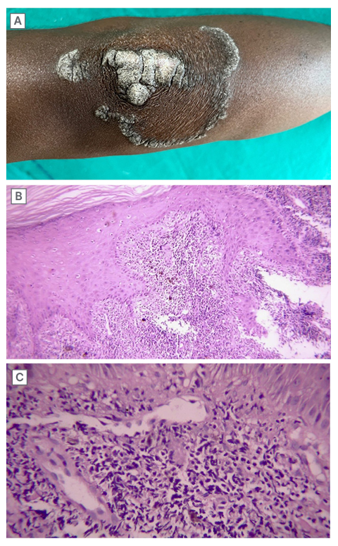
Figure 1: Figures showing clinical and histopathological images of the presented case.
A) Non-tender verrucous annular plaque over the left elbow with some scaling and dyspigmentation. B) 10x magnification showing hyperkeratosis without any parakeratosis, wedge-shaped hypergranulosis, saw-tooth rete ridges, vacuolar degeneration of the basal cell layer, thickening of the basement membrane, and lymphocytic infiltrates at the dermo-epidermal junction. C) 40x magnification showing perivascular and peri-adnexal lymphocytic infiltrates, pigment incontinence, and mucin deposition in the dermis.
DIF was performed on perilesional skin, which showed granular deposition of IgG and complement component 3 (C3) along the dermo-epidermal junction (lupus band) together with linear fibrinogen deposition along the basement membrane zone, a pattern characteristic of LP.7
Serological examinations demonstrated a positive ANA test with a speckled pattern at a titre of 1:160. Anti-double-stranded DNA antibodies and extractable nuclear antigen profile were negative. Complete blood count, hepatic enzymes, and renal function tests, urinalysis, and chest radiography were within normal limits.
Based on combined clinical findings, histopathological overlap, dual DIF pattern, and serological positivity, a diagnosis of cutaneous-limited LE–LP overlap syndrome was established. According to the criteria proposed by Lee et al., the case fulfilled criteria for ‘possible’ overlap syndrome.2
The patient was treated with topical clobetasol propionate 0.05% ointment applied twice daily for 3 weeks, along with emollients and photoprotection advice. On follow-up, there was significant reduction in pruritus, marked flattening of the plaque, and reduction in hyperkeratosis, with residual post-inflammatory dyspigmentation. The patient continues to be followed for recurrence or evolution of systemic disease.
DISCUSSION
LE–LP overlap syndrome occupies a unique position within the spectrum of interface dermatoses, representing a convergence of two well-characterised autoimmune diseases. Its rarity, protean manifestations, and histological overlap with pure LE or LP contribute to frequent diagnostic delay or misclassification.1,2
Clinical Spectrum and Diagnostic Pitfalls
The clinical presentation of LE–LP overlap syndrome is highly variable. Lesions may resemble classical LP or discoid LE, or display hybrid morphology with annular configuration, violaceous hue, scaling, dyspigmentation, or atrophy (Table 1).2,11
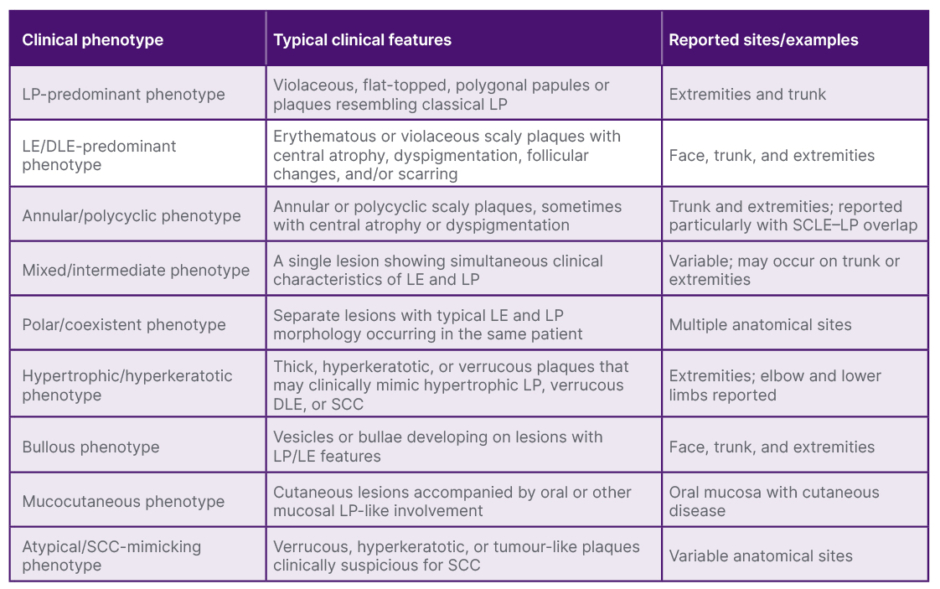
Table 1: Spectrum of reported clinical presentations of LE–LP overlap syndrome.
DLE: discoid lupus erythematosus; LE: lupus erythematosus; LP: lichen planus; SCC: squamous cell carcinoma; SCLE: subacute cutaneous lupus erythematosus.
Pathogenesis
The immunopathogenesis of LE–LP overlap syndrome likely reflects shared immune pathways rather than coincidental coexistence. Both LE and LP are characterised by CD8+ T cell-mediated keratinocyte injury and heightened Type I IFN signalling.3,5 Plasmacytoid dendritic cells and IFN-α play central roles in sustaining interface inflammation in both diseases.13 Drug-induced immune modulation has also been implicated, with reports of overlap syndrome arising in association with the usage of aromatase inhibitors and malignancy-related immune dysregulation.5
Management
Management strategies depend upon the disease extent and the severity. Localised cutaneous disease often responds well to potent topical or intralesional corticosteroids, or topical calcineurin inhibitors.14 More extensive or hypertrophic lesions may require systemic therapy, including antimalarials such as hydroxychloroquine, or retinoids such as acitretin, or immunosuppressive agents.10,15 Recent advances in understanding IFN-driven pathways raise the possibility of targeted biologic therapies in refractory cases.5
Differentiation from HLP and Long-Term Outcomes
HLP is an important clinical and histopathological differential diagnosis in verrucous or hypertrophic lesions of LE–LP overlap syndrome. HLP typically presents as intensely pruritic, hyperkeratotic, or verrucous plaques, often affecting the distal lower extremities, particularly the shins, and may be bilateral or multifocal. In contrast, LE–LP overlap syndrome is uncommon and more frequently demonstrates mixed clinical features of LE and LP, with annular, violaceous, discoid, or atrophic morphology and a tendency to involve photo-exposed sites, although atypical verrucous lesions at non-sun-exposed sites may occur, as in this patient. Histopathologically, both conditions may demonstrate hyperkeratosis, hypergranulosis, saw-toothed acanthosis, basal vacuolar degeneration, and a lichenoid infiltrate. However, wedge-shaped hypergranulosis, hyperorthokeratosis, and irregular psoriasiform epidermal hyperplasia are characteristic supportive features of HLP.16,17 In contrast, the presence of prominent basal vacuolar degeneration, basement membrane thickening, peri-adnexal/perivascular inflammation, and dermal mucin favours LE. In the present case, although wedge-shaped hypergranulosis and saw-toothed rete ridges suggested LP/HLP, the associated dermal mucin, prominent peri-adnexal inflammation, positive ANA, and dual DIF pattern provided additional evidence for LE–LP overlap syndrome. The 2025 review by Scotti et al.16 also emphasises that adequate biopsy depth and clinicopathological correlation are particularly important in distinguishing hypertrophic lichenoid disorders from hypertrophic lupus and SCC.
The two conditions may also differ in their long-term clinical implications. HLP is frequently chronic and therapeutically challenging when extensive, and longstanding hypertrophic lesions warrant surveillance because SCC has been reported to arise within HLP, particularly in lesions of the lower extremities.16,18 LE–LP overlap syndrome, in contrast, has a less well-defined natural history because of its rarity and limited long-term follow-up data. The principal concerns are persistence or recurrence of cutaneous disease and, in selected patients, subsequent development of systemic lupus manifestations. Therefore, continued clinical surveillance is appropriate, particularly when lesions are chronic, hypertrophic, ulcerated, rapidly enlarging, or otherwise clinically suspicious for malignant transformation. However, LP and LE can also coexist in the same patient. Blaschkoid LP in the setting of concurrent childhood-onset systemic LE has been described in literature.19
Malignant Transformation in Chronic Cutaneous Lupus
Chronic cutaneous LE, particularly longstanding discoid LE (DLE), is associated with an increased risk of cutaneous SCC, although the absolute risk remains relatively low and estimates vary substantially among studies. Traditionally, the risk has been cited at approximately 2–5%, while some older literature and selected series have reported substantially higher figures, approaching 10–15%, particularly in longstanding, hypertrophic, or extensively scarred lesions.20-22 More recent systematic and population-based data suggest that SCC arising within DLE remains an uncommon complication, with reported rates generally lower than the highest historical estimates.21,22 The risk appears to be influenced by chronic inflammation and scarring, cumulative UV exposure, lesion duration, and anatomical site. SCC arising in DLE may also behave more aggressively than conventional cutaneous SCC, with recurrence and metastatic disease reported in the literature.21 Consequently, persistent induration, ulceration, crusting, bleeding, pain, or rapid enlargement of a chronic DLE lesion should prompt biopsy to exclude malignant transformation. Although this patient did not have DLE or evidence of malignancy, recognition of this risk is relevant when considering the long-term implications of lupus-associated hypertrophic or verrucous lesions.
CONCLUSION
LE–LP overlap syndrome is a rare but important diagnostic consideration in patients presenting with chronic, atypical lichenoid or verrucous plaques. This case highlights an unusual presentation on a non-sun-exposed site, emphasising the need for high clinical suspicion and comprehensive evaluation. Histopathology, direct immunofluorescence, and serological evaluation are the cornerstones for accurate diagnosis. Early recognition enables clinicians to manage appropriately, avoid misdiagnosis, and facilitate long-term monitoring for systemic involvement.