Abstract
Menopause is a natural biological transition characterised by declining oestrogen levels, with significant effects on skin structure, barrier function, and inflammation. While these physiological changes are well recognised, the impact of menopause on common inflammatory dermatoses remains poorly understood. This review examines the current evidence on acne, rosacea, hidradenitis suppurativa, psoriasis, lichen sclerosus, and lichen planus in peri- and postmenopausal women, focusing on clinical presentation and management. The relationship between menopause and individual inflammatory dermatoses is complex, with considerable variation in disease presentation and response to hormonal treatment. In particular, the role of hormone replacement therapy remains uncertain. Management should therefore continue to follow established disease-specific guidelines, while taking into account menopausal symptoms, hormonal treatments, skin sensitivity and associated comorbidities. Further prospective studies incorporating hormonal measurements, molecular markers, and treatment responses are needed to better understand the relationship between menopause and inflammatory skin disease and develop evidence-based management strategies for women during and after the menopausal transition.
Key Points
1. Menopausal hormonal changes influence skin structure, barrier function, and inflammation, yet their effects on common inflammatory dermatoses remain poorly understood, presenting challenges for clinicians managing women during this transition.2. This review examines current evidence on acne, rosacea, hidradenitis suppurativa, psoriasis, lichen sclerosus, and lichen planus in peri- and postmenopausal women, focusing on clinical presentation and management considerations.
3. In the absence of menopause-specific treatment guidelines, clinicians should follow established disease-specific recommendations while considering menopausal symptoms, hormone replacement therapy, and associated comorbidities, adopting a holistic, multidisciplinary approach where appropriate.
INTRODUCTION
Menopause is a natural biological transition characterised by the permanent cessation of menstruation and declining ovarian oestrogen and progesterone production, typically occurring between 45–55 years of age.1,2 Skin ageing is a multifactorial process influenced by intrinsic factors, including genetics and hormonal changes, and extrinsic factors, including photodamage, smoking, and environmental stressors.3 Oestrogen plays a key role in maintaining skin homeostasis by regulating collagen synthesis, hydration, elasticity, and epidermal barrier function.2 Consequently, oestrogen deficiency contributes to significant collagen loss and reduced skin elasticity, resulting in reduced skin thickness and wrinkling, alongside xerosis and impaired wound healing,2,4 which can significantly impact the quality of life.5,6
Although the effects of menopause on physiological skin ageing are well recognised, its influence on inflammatory dermatoses has received comparatively little attention. Given the role of oestrogen in skin homeostasis and modulation of inflammation, hormonal changes during menopause may also influence inflammatory dermatoses.1 Emerging evidence suggests that menopause may alter the presentation and clinical course of several skin conditions, although the strength of evidence varies considerably between conditions.7 In a survey of women attending a menopause clinic, almost half reported worsening of a pre-existing skin condition following menopause,6 emphasising the importance of considering menopause when assessing women with inflammatory dermatoses.
Women now spend almost one-third of their lives after menopause,8 making an understanding of menopause-associated inflammatory dermatoses increasingly relevant to dermatological practice. This review summarises the current evidence surrounding common inflammatory dermatoses during menopause, discusses practical considerations for diagnosis and management, and highlights priorities for future research.
ACNE
Acne is well recognised in perimenopause, affecting approximately 25% of women.4 In later life, it remains more common in women than men, with one study reporting acne in 26.3% of women aged 40–49 years compared with 12.0% of men.9 Menopausal acne may present as persistent, recurrent, or new-onset disease, with persistent acne being the most common, accounting for approximately 75–85% of adult female acne compared with 20–40% for late-onset disease.10 Although prevalence declines with advancing age, acne in midlife remains clinically important because it may result in permanent scarring and significant psychosocial morbidity.4,10
The pathogenesis of menopausal acne is incompletely understood. Oestrogen levels decline rapidly following menopause, while androgen levels decrease more gradually, resulting in a relative androgen excess that may contribute to acne development.10 Other contributing factors may include abnormal follicular hyperkeratinisation, altered sebum production, chronic low-grade inflammation, impaired epidermal barrier function, alterations in the cutaneous microbiome, and metabolic and stress-related pathways.10,11
Clinical assessment should take into account both the physiological changes associated with oestrogen-deficient skin and the potential contribution of hormonal factors. The presence of androgenic alopecia or virilisation should prompt further evaluation for underlying causes of hyperandrogenism.10
Management of menopausal acne can be challenging, as post-inflammatory hyperpigmentation and scarring from previous episodes may become more apparent with age, while increased skin dryness and sensitivity may limit treatment tolerability.10
Maintaining skin barrier function is important, and skincare routines should include gentle cleansers and regular moisturisers that support the skin barrier and microbiome.12 Impaired barrier function may increase inflammation and exacerbate acne.12
Topical retinoids are a mainstay of acne treatment and are useful for both active disease and long-term maintenance. Azelaic acid is effective for both inflammatory and non-inflammatory acne and may be particularly useful where post-inflammatory hyperpigmentation is present.4,10,11,13
Procedural treatments, including chemical peels and laser therapies, may also have a role in menopausal acne management.10 Laser and light-based therapies may be useful adjuncts, particularly for inflammatory acne, through effects on inflammation, sebaceous gland activity, and Cutibacterium acnes. Various modalities, including pulsed dye lasers, have been used for inflammatory acne and may also improve acne scarring; however, their role specifically in menopausal acne requires further investigation.10
Antiandrogen treatments have been investigated in late persistent female acne, with spironolactone demonstrating an improvement in acne at 6 months.14 Spironolactone may be considered in appropriately selected postmenopausal women.4,10,11 The SAFA trial also reported lower oral antibiotic use among women allocated to spironolactone at follow-up of up to 52 weeks, although participant numbers were small.14 Topical clascoterone 1%, an androgen receptor inhibitor, represents a further treatment option for acne.15 However, the efficacy of these anti-androgen treatments specifically in menopausal women has not been established.14,15
Hormonal therapies should also be reviewed, as androgenic progestogens such as levonorgestrel may contribute to acne.11 Whether switching to a less androgenic alternative, such as micronised progesterone,16 has any benefit for acne remains to be explored. Clinicians should also consider exogenous testosterone, prescribed for hypoactive sexual desire disorder, as a potential contributor to new-onset or worsening acne.7
Future prospective studies are needed to better define the mechanisms underlying menopausal acne and inform management specifically for peri- and postmenopausal women.
ROSACEA
Rosacea is a common chronic inflammatory dermatosis that is particularly prevalent in women.1 Although the relationship between menopause and rosacea remains poorly understood, menopausal flushing is recognised to precipitate or exacerbate disease activity.1,4,8
The mechanisms linking menopause and rosacea are likely multifactorial. Declining oestrogen levels may impair epidermal barrier function and increase cutaneous vasoreactivity and skin sensitivity, potentially exacerbating rosacea-associated inflammation.17 Menopause-related hormonal changes may also alter the cutaneous microbiome, with a possible increase in Demodex folliculorum proposed as a contributor to rosacea exacerbation, although direct evidence for this menopause-specific mechanism remains limited.17
Despite these proposed mechanisms, clinical evidence examining the relationship between menopause and rosacea remains inconsistent.7 A small prospective study proposed an ‘oestradiol protective hypothesis’ after demonstrating an association between lower circulating oestradiol levels and greater rosacea severity.18 However, this hypothesis is challenged by a substantially larger cohort study demonstrating a lower incidence of rosacea after menopause despite declining endogenous oestrogen levels, together with an increased risk among women receiving hormone replacement therapy (HRT).19 Collectively, these findings suggest that the relationship between endogenous hormones, exogenous hormone therapy, and rosacea is likely more complex than can be explained by oestrogen deficiency alone.
Management should continue to follow established rosacea guidelines. Clinicians should recognise menopausal flushing as a potential trigger for disease exacerbation. Menopausal status and HRT exposure should also be considered when evaluating women presenting with new-onset or worsening rosacea, given the reported association between HRT use and increased rosacea risk.7,19 Daily broad-spectrum UVA and UVB photoprotection and a gentle skincare routine incorporating appropriate cleansers and moisturisers should also be encouraged, particularly in women with increased skin sensitivity.20 Topical azelaic acid may be considered for inflammatory lesions, introduced gradually to minimise irritation in patients with sensitive skin. Vascular laser or intense pulsed light therapy may also be considered for persistent erythema and telangiectasia.20
HIDRADENITIS SUPPURATIVA
Hidradenitis suppurativa (HS) is a chronic inflammatory dermatosis in which hormonal factors are thought to contribute to disease activity.1 Clinical studies have reported variable effects of menopause on HS, with improvement, persistence, and worsening of disease all described.21,22 In a survey by Fernandez et al.,21 39.5% of women reported worsening of HS symptoms following menopause, while 44.2% reported no change.21 These findings highlight the heterogeneous effect of menopause on HS disease activity.
Disease activity commonly fluctuates with the menstrual cycle,23 and HS is frequently associated with acne and menstrual irregularity, supporting a role for sex hormones in disease expression.22 Evidence from hormonal therapies provides further support for hormonal involvement, with clinical improvement reported following treatment with combined oral contraceptives and other hormone-modulating therapies.23,24 However, the mechanisms underlying these observations remain incompletely understood. An immunohistochemical study found no significant differences in androgen or oestrogen receptor expression within HS lesions.25 The role of HRT in HS remains uncertain, with evidence regarding its use currently limited to anecdotal reports.23
Endocrine and metabolic comorbidities, including obesity and insulin resistance, may also contribute to HS disease burden.26 As both HS and menopause are independently associated with increased cardiometabolic risk, assessment and optimisation of these risk factors should form part of routine care, with multidisciplinary input from dermatology, endocrinology, and other relevant specialties where appropriate.7,26,27
Management should continue to follow established HS guidelines. Given the potential influence of hormonal factors, clinicians should consider menopausal status and HRT when evaluating women with changing disease activity.7,23,24
PSORIASIS
Psoriasis is a chronic, immune-mediated inflammatory dermatosis occurring in genetically susceptible individuals, with disease activity influenced by hormonal changes.28,29 Clinical improvement is commonly observed during pregnancy, whereas many women report worsening symptoms following menopause.28 However, epidemiological studies examining the impact of menopause on psoriasis have produced conflicting findings, and the overall influence of menopause on disease activity remains uncertain.7
Declining oestrogen levels have been proposed to contribute to psoriasis exacerbation through effects on Th1-mediated immune responses.29 However, this hypothesis has not been consistently supported by clinical studies, and the mechanisms linking menopause and psoriasis remain incompletely understood.7
Whether this hormonal influence translates into a therapeutic role for HRT remains uncertain, and many existing studies investigated formulations that are less commonly used in current clinical practice.7 Observational studies have reported conflicting findings, with some demonstrating an increased risk of psoriasis among HRT users, others reporting no significant association, and one suggesting a reduced risk with combined oestrogen-progestogen therapy.30 Collectively, these findings suggest that responses to HRT may vary between individuals and according to HRT formulation, and current evidence is insufficient to support its use for psoriasis management.7
Beyond disease activity, women with psoriasis should also undergo assessment and optimisation of cardiometabolic risk factors, as both psoriasis and menopause are independently associated with an increased risk of cardiovascular disease, metabolic syndrome, and depression.31
Management should continue to follow established psoriasis guidelines. Women commencing HRT should be monitored for changes in psoriasis activity, with management individualised according to clinical response.7
LICHEN SCLEROSUS AND GENITOURINARY SYNDROME OF MENOPAUSE
Lichen sclerosus (LS) is a chronic autoimmune inflammatory scarring dermatosis that predominantly affects the anogenital region and is most commonly diagnosed in postmenopausal women.32
Genitourinary syndrome of menopause (GSM) describes a constellation of genital and urinary symptoms and signs resulting from oestrogen deficiency, including vaginal dryness, soreness, burning, itching, and urinary symptoms.32 Symptoms may overlap with those of LS, and both conditions may coexist, making accurate diagnosis particularly important in postmenopausal women.32 Early recognition is important, as GSM symptoms may worsen over time if untreated.32 This distinction is also clinically important because untreated LS may result in progressive scarring, anatomical distortion, and an increased risk of vulval squamous cell carcinoma.32,33
Assessment should include enquiry about symptoms of GSM, including urinary and vaginal symptoms, to ensure that coexistent disease is not missed. Management should continue to follow established LS guidelines irrespective of menopausal status. High-potency topical corticosteroids remain the first-line treatment, with long-term follow-up recommended to monitor treatment response and facilitate early detection of malignant transformation.32-34 The effect of HRT on the clinical course of LS has not been examined.7,32 However, in women with coexistent GSM, early treatment with local vaginal oestrogen may improve symptoms attributable to oestrogen deficiency but does not replace standard anti-inflammatory treatment for LS.32 A recent retrospective study of women with vulvar LS reported that vulvar intraepithelial neoplasia and invasive vulvar neoplasia occurred predominantly in women over 50 years of age, reinforcing the importance of ongoing surveillance in this population.34
LICHEN PLANUS
Lichen planus (LP) is a chronic immune-mediated inflammatory disorder affecting the skin and mucous membranes.32 Vulvovaginal LP most commonly presents in peri- and postmenopausal women and remains an important differential diagnosis to LS, as symptoms of the two conditions may overlap.32 Oral LP is also more prevalent in women over 40 years of age, with one study reporting a higher prevalence in perimenopausal than premenopausal women.35,36
The relationship between menopause and LP remains poorly understood, although hormonal changes have been proposed to influence disease development.36 Management should continue to follow established treatment guidelines, with topical corticosteroids remaining first-line therapy.36 The role of HRT in oral LP remains uncertain, and further studies are required to clarify its influence on disease activity.
Key menopause-related considerations and practical management points for the inflammatory dermatoses discussed are summarised in Table 1.
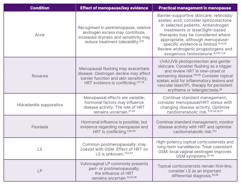
Table 1: Menopause-related considerations and practical management of inflammatory dermatoses.
GSM: genitourinary syndrome of menopause; HRT: hormone replacement therapy; IPL: intense pulsed light; LP: lichen planus; LS: lichen sclerosus.
CONCLUSION AND FUTURE DIRECTIONS
Menopause represents an important physiological transition that may influence the presentation, severity, and management of several inflammatory dermatoses. Although hormonal changes appear to contribute to disease activity in selected conditions, the evidence base remains limited, and menopause-specific management recommendations are lacking for most inflammatory skin diseases.
Management should continue to follow established disease-specific guidelines while recognising the potential influence of menopausal symptoms and HRT in midlife women. Given the shared burden of systemic comorbidities in conditions such as HS and psoriasis, clinicians should adopt a holistic approach that includes optimisation of cardiometabolic health and collaboration with primary care and menopause specialists where appropriate.
Further prospective studies incorporating measurements of hormone levels and relevant molecular markers are needed to clarify the effects of endogenous hormonal changes and HRT on disease susceptibility, severity, and treatment response. Future studies should also evaluate currently prescribed HRT formulations, as much of the existing evidence is based on formulations that are less commonly used in current clinical practice. Improving our understanding of these interactions may facilitate the development of evidence-based menopause-specific management strategies and support more personalised dermatological care for women during the menopausal transition.