Abstract
Introduction: Klippel–Trenaunay syndrome (KTS) is an extremely rare congenital disorder belonging to the wider group of PIK3CA-related overgrowth spectrum disorders, and it is characterized by vascular malformations. It usually presents with a clinical triad of port-wine stains, soft tissue and bone hypertrophy, and vascular malformations mainly affecting the lower limbs. Its etiology has been linked to sporadic mutations in the PIK3CA gene. This case contributes to the limited body of literature from South Asia and highlights regional presentation and diagnostic challenges due to its rarity. Although port-wine stains are classically described in KTS, the presence of multiple widespread cutaneous hemangiomas has not been widely documented in association with this syndrome.
Case presentation: A 4-year-old girl presented with multiple blue-colored, tortuous swellings in the right lower limb, including the ventral lateral aspect of the foot, knee, labia majora, and posterior thigh. The swellings were irregular in shape, compressible, non-tender, and demonstrated minimal flow on color Doppler examination. Sequential progression was noted from foot to buttock. A port-wine stain was observed on the buttocks. Additionally, several hemangiomas were scattered throughout body regions including the axilla, left leg, left arm, and post-auricular area. Some lesions were bleeding and crusted. Developmental milestones were achieved appropriately for age.
Conclusion: The diagnosis of KTS was established based on clinical findings and imaging investigations including color Doppler ultrasound, CT angiography, and MRI. Imaging demonstrated venolymphatic malformations with absence of arterial feeders. Conservative management was adopted. The patient remained clinically stable without major complications during follow-up. This case expands the dermatological spectrum of KTS by describing multiple widespread hemangiomas in association with classic KTS features. Imaging plays a pivotal role in differentiating KTS from overlapping vascular syndromes such as Parkes Weber and Sturge–Weber syndrome, especially in resource-limited settings.
Key Points
1. Klippel–Trenaunay syndrome (KTS) can present with atypical, widespread, cutaneous hemangiomas in addition to its classical triad, expanding its known dermatological spectrum.2. Multimodal imaging (Doppler ultrasound, CT angiography, and MRI) is essential for confirming low-flow venolymphatic malformations and differentiating KTS from overlapping syndromes such as Parkes–Weber syndrome and Sturge–Weber syndrome.
3. Conservative management with close follow-up remains a practical and effective approach in pediatric KTS, particularly in resource-limited settings
INTRODUCTION
Klippel–Trenaunay syndrome (KTS) is a rare congenital vascular malformation syndrome characterized by capillary malformations, venous abnormalities, and limb hypertrophy. Its etiology has been linked to sporadic somatic mutations in the PIK3CA gene. The diagnosis is established when at least two components of the classical triad are present. PIK3CA-related overgrowth spectrum (PROS) disorders demonstrate marked clinical heterogeneity due to postzygotic mosaic PIK3CA mutations, which may result in a wide phenotypic spectrum ranging from isolated localized overgrowth to complex multi-tissue involvement.1
Although the International Society for the Study of Vascular Anomalies (ISSVA) 2018 classification remains the standard framework for the classification of vascular anomalies, it primarily emphasizes vascular morphology. Recent advances in molecular biology have led to the development of more comprehensive classification systems that incorporate genetic and phenotypic features. In particular, the classification proposed by Canaud et al.2 integrates vascular malformations with segmental overgrowth and underlying PIK3CA mutations, providing a more unified understanding of these disorders. Within this framework, KTS is considered part of the PROS, which encompasses a spectrum of disorders characterized by congenital or early-onset segmental overgrowth and associated vascular anomalies. This expanded classification has important implications for diagnosis and future targeted therapies, particularly in complex or atypical presentations.
Distinguishing KTS from Parkes–Weber syndrome (PWS) is essential and relies on identifying the absence of arteriovenous malformations or fistulas.3 Capillary malformations, venous abnormalities, and limb overgrowth have been reported with incidence rates of 98%, 72%, and 67%, respectively, in a study by the Mayo Clinic.3 Elevated D-dimer levels have been shown to be highly specific for venous malformations.4
Although visceral hemangiomas, including bladder, splenic, gastrointestinal, and intraneural hemangiomas, have been described in KTS,5-8 widespread cutaneous hemangiomas are rarely reported in association with this syndrome, with limited literature available from South Asia. This case may further expand the recognized dermatologic spectrum of KTS.
PATIENT INFORMATION
The patient was a 4-year-old female child from Pakistan. All identifying details have been omitted to preserve confidentiality. She was born full-term without antenatal or perinatal complications.
Primary Concerns and Symptoms
The earliest manifestation was a bruise-like discoloration on the plantar surface of the right foot noted at birth. Over time, this evolved into nodular swellings. The child developed multiple, blue-colored tortuous swellings extending from the foot to the buttocks region. A port-wine stain was observed on the buttocks. Subsequently, raised red-purple vascular lesions began appearing over the axilla, chest wall, extremities, and behind the auricular region. Some lesions demonstrated crusting and occasional bleeding (Figure 1A-D). Developmental milestones were achieved appropriately for age.
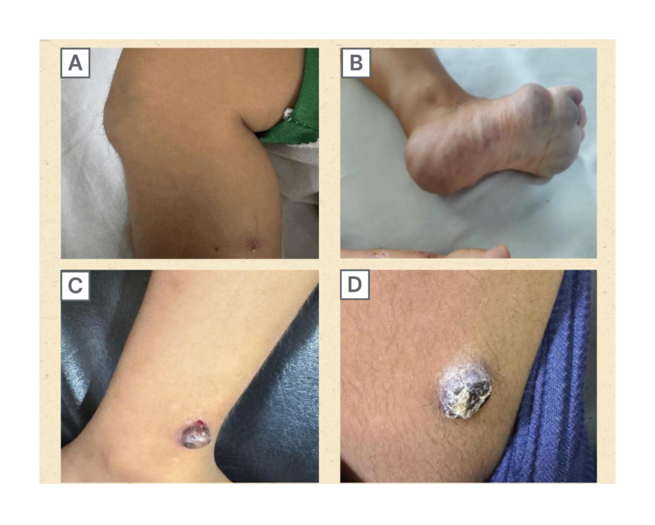
Figure 1: Hemangiomas.
Figure 1A shows a knee swelling. Figure 1B shows a blue-colored tortuous swelling on the right foot (ventral lateral side). Figure 1C shows a bleeding hemangioma on anterior aspect of the lower leg just above the ankle (pink-purple). Figure 1D shows a calcified hemangioma on the left thigh.
Medical, Family, and Psychosocial History
There was no known family history of vascular malformations or genetic disorders. No psychosocial stressors were reported. There was no clinical history suggestive of overlapping neurocutaneous syndromes.
Relevant Past Interventions
No prior surgical or invasive interventions had been performed before the presentation.
Physical Examination and Important Clinical Findings
Clinical examination revealed intact sensations, normal pulses, and no audible bruit on auscultation. The port-wine stain on the buttocks appeared flat, non-pulsatile, and non-blanching. Swellings decreased at rest and increased with movement reflex. Femoral artery, dorsalis pedis, and posterior tibialis were intact. Several dome-shaped, well-defined, dark red-to-purple vascular lesions were observed over the axilla, left leg, left arm, chest wall, and post-auricular region. Some lesions were lobulated, crusted, calcified, and occasionally bleeding. Sensations were intact, and peripheral pulses, including femoral artery, dorsalis pedis, and posterior tibialis, were normal.
TIMELINE
At birth, a bruise-like lesion was noted on the right plantar aspect of the foot. During infancy, nodular swellings progressively developed along the right lower limb. Over the subsequent years, multiple raised vascular lesions appeared over different body regions.
At 4 years of age, comprehensive imaging studies were performed, leading to diagnostic evaluation. The patient first presented to the orthopedic outpatient department (Month 0) when the mother expressed concern regarding progressive skin and soft tissue lesions. Clinical examination was performed at presentation, followed by radiological investigations, including Doppler ultrasound, CT/MRI legs, and baseline laboratory workup including complete blood count, urine routine examination, renal function tests, and liver function tests within the next month (i.e., Month 1).
In Month 5, a more detailed systemic evaluation was performed in view of reported associations of KTS with visceral involvement in the literature, as well as to exclude potential internal hemangiomas or occult bleeding. This included X-Ray kidney, ureter, and bladder; abdominal ultrasonography;8 chest radiography;9 and renal function tests, all of which were within normal limits.
A neurological evaluation was also sought. An EEG conducted in Month 2 was reviewed and was considered likely artifactual by the consulting neurologist. Although the EEG report suggested multifocal epileptiform activity, the patient had no corresponding clinical seizure manifestations. Neuroimaging was discussed; however, brain MRI was not performed as it was not available in the clinical setup. In addition, given the absence of neurological symptoms and the specialist opinion that further imaging was unlikely to alter management, a decision was made to defer neuroimaging following a risk–benefit assessment in a pediatric patient.
Following this assessment, the patient continued regular follow-up in the dermatology clinic, where multiple vascular skin lesions were managed with serial cryotherapy sessions (cryoablation).
The patient was subsequently followed from Month 5 up to 2 years with multidisciplinary involvement, including cardiology, dermatology, and internal medicine, for ongoing assessment and management.
DIAGNOSTIC ASSESSMENT
Initial laboratory investigations revealed low hemoglobin levels and significantly elevated D-dimer levels. Doppler ultrasound demonstrated minimal flow within the lesions and supported venous and lymphatic malformations. MRI revealed extensive abnormal tubular and serpiginous enhancing structures involving the right lower limb from the buttock to the ankle, extending into subcutaneous tissues and muscle compartments, indicating venous malformations.
CT angiography demonstrated scattered soft tissue density in the right gluteal region, perineum, and lower extremity. A dilated tortuous lateral marginal vein was identified. Hypoplastic and intact great saphenous veins were visualized. No definite arterial feeders were identified.10
IMAGING STUDIES
MRI
Contrast-enhanced multiplanar MRI revealed extensive tubular and serpiginous vascular abnormalities involving the right lower limb, with continuous extension from the gluteal region through the thigh, knee, leg, and ankle (Figure 2A) The lesions showed marked post-contrast enhancement within muscular compartments (Figure 2A) with additional extension into the subcutaneous tissues (Figure 2B) consistent with widespread soft-tissue vascular involvement. The abnormal structures are not limited to the skin surface but extend into deeper layers, specifically the subcutaneous tissue and the muscle compartments of the right lower limb, indicating it to be venous malformation.11
Venous malformations have variable presentations ranging from small, localized, superficial lesions to large, infiltrative lesions with poorly defined margins that cross soft tissue planes.11
The presence of abnormal structures is associated with swelling in the right lower limb as seen on MRI, suggesting a potential impact on fluid dynamics or lymphatic drainage in the affected areas. Lymphatic malformations were also supported by the Doppler ultrasound report.
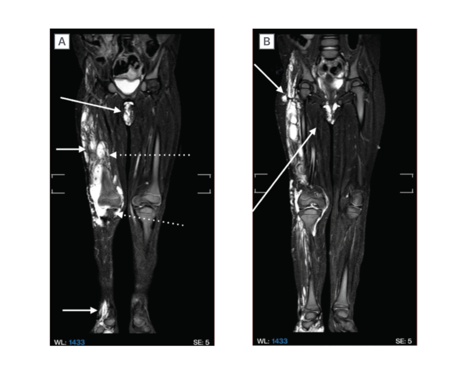
Figure 2: MRI lower legs.
Multiplanar contrast MRI showing extensive abnormal tubular/serpiginous structures involving the right lower limb, including the buttocks region, inferior thigh region, knee region, leg and ankle (arrows). It shows post-contrast enhancement in subcutaneous tissue (arrows) and involvement of muscular compartments (dotted arrows).
CT Angiogram
In this case, a bruise-like lesion was noted on the right plantar aspect of the foot with CT angiogram at birth. During infancy, nodular swellings progressively developed along the right lower limb.
Over the subsequent years, multiple raised vascular lesions appeared over different body regions.
Findings show venous abnormalities in the right lower limb, gluteal, and perineal regions.
Imaging demonstrated a hypoplastic great saphenous vein on the affected side, while the contralateral great saphenous vein appeared intact and of normal caliber. Additionally, multiple tortuous dilated vascular channels were identified along the dorsal and lateral aspects of the right foot, extending proximally into the leg and gluteal region (Figure 3A and 3B).
Proliferation of synovial vessels is also observed at the level of the knee joint. Dilated superficial venous channels in the buttock and perineum are mainly draining into the ipsilateral internal iliac vein.
A dilated and tortuous vessel, indicative of a lateral marginal vein, was also observed (Figure 3C) This vein is typically an embryonic remnant of part of the superficial venous system and is often associated with KTS.12 Lateral marginal veins and patent sciatic veins are seen in 72% of patients with KTS.4
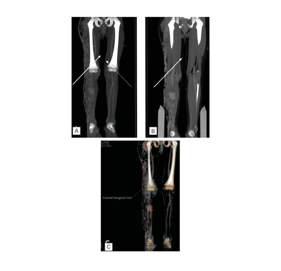
Figure 3: CT angiogram legs.
Arrows show the small calibre (hypoplastic) great saphenous vein and dotted arrows show the intact great saphenous vein. The image also shows tortuous dilated vessels in the right foot dorsal and lateral aspects, extending upwards into the leg and gluteal region. Proliferation of synovial vessels is also observed at the level of the knee joint. In this case, CT angiography findings show venous abnormalities in right lower limb, gluteal, and perineal regions. The image also shows patent lateral marginal vein.
Multiple scattered areas of abnormal tissue density or nodular foci are observed, with one notable lesion measuring 51×35 mm in the buttock region. Additionally, some of these foci show internal calcifications.
Histopathological confirmation was not pursued due to concerns regarding wound care, increased risk of infection, and potential sepsis in a pediatric patient within a developing country setting.
Despite the presence of vascular anomalies, the imaging studies did not reveal any definitive arterial feeders contributing to these malformations. This suggests that the observed abnormalities primarily involve the venous system rather than arterial vessels.
The diagnosis of KTS was established based on the presence of capillary malformation, venous abnormalities, and limb hypertrophy. PWS was ruled out due to the absence of arteriovenous malformations. Sturge–Weber syndrome (SWS) was considered because EEG showed epileptic waveforms; however, clinical history and examination were insufficient for diagnosis, and further neuroimaging was recommended.
The prognosis remains favorable with conservative management, although long-term monitoring is necessary for potential complications, including thrombosis, bleeding, and possible internal organ involvement.
THERAPEUTIC INTERVENTION, FOLLOW-UP, AND OUTCOMES
The patient remained clinically stable without progression of limb hypertrophy or systemic complications. Conservative management was well tolerated.
Although pharmacological and interventional options are described in the literature for similar rare vascular overgrowth disorders, management remains largely non-standardized. As highlighted by Khandelwal et al.,13 conservative management is often preferred due to the rarity of the condition, limited evidence, and clinician caution regarding invasive or drug-based therapies. This approach is especially relevant in resource-limited settings, where conservative care becomes the most practical and safe strategy.
The management of the authors’ patient was primarily directed toward symptomatic cutaneous lesions. Specifically, episodes of bleeding from the vascular skin lesions were managed with local care, and the patient underwent serial cryotherapy sessions (cryoablation) for treatment of the hemangiomas. In addition, the patient’s guardians were counseled regarding proper hygiene and local care of the lesions, including keeping the affected areas clean to reduce the risk of secondary infection and bleeding. Emphasis was placed on the importance of monitoring disease progression during growth and recognizing warning signs warranting earlier medical review, including new-onset pain, swelling, redness, or functional impairment.
DISCUSSION
KTS is a rare congenital vascular disorder characterized by vascular malformations involving capillary, venous, and sometimes lymphatic vessels, and is often associated with hypertrophy of soft tissues and bones. Lymphatic anomalous development may also occur due to tissue overgrowth caused by somatic mutations.14 The presence of two of these three signs is enough to obtain the diagnosis. According to the vascular anomaly classification proposed by Wassef et al.,14 KTS is categorized under combined vascular malformations of the capillary-lymphatic-venous malformation type.2
This case report presents a 4-year-old girl diagnosed with KTS. The patient has several venous lymphatic abnormalities on her right leg along with a patent and tortuous lateral marginal vein. External genital involvement, although seldom reported, was observed in the authors’ patient; a similar manifestation has been described by Mathur et al.15 in a 3-year-old male child with penoscrotal involvement, suggesting that such rare presentations can occur in both sexes.
In addition, involvement of the genitourinary and gastrointestinal tract has been reported in over 30% of patients with KTS, affecting organs such as the bladder, urethra, uterus, and external genitalia.16 In the authors’ case, physical examination of the perineum and MRI findings demonstrated vulvar involvement; however, hematuria was not observed.
Dermatological manifestations were also present, notably a non-blanching port-wine stain located on the right buttock. This stain, consistent with capillary malformations, is red-to-purple, remains within a dermatomal distribution, and does not cross the body’s midline, as previously documented by Zakko et al.17 These findings align with common characteristics of KTS. In a study from the Mayo Clinic, Jacob et al.3 reported that capillary malformations, venous-lymphatic abnormalities, and limb hypertrophy occur in approximately 98%, 72%, and 67% of patients, respectively. The symptoms of frequent rash flares and elevated body temperature were also noted in the authors’ patient.
Elevated D-dimer levels were reported in the authors’ case, which is significant because increased D-dimer may indicate ongoing coagulation and fibrinolysis associated with venous malformations, a common feature in KTS. Such elevations can reflect localized intravascular coagulation within abnormal vascular channels and may help identify patients at increased risk for thrombotic complications such as deep vein thrombosis or pulmonary embolism. The diagnostic value of D-dimer in vascular anomalies has been described by Dompmartin A et al.,4 and patients with KTS may develop complications, including deep vein thrombosis, pulmonary embolism, bleeding, and orthopedic abnormalities due to underlying vascular malformations and abnormal venous circulation, as reported by Amir et al.18
The term ‘hemangioma’ has been used in various contexts, sometimes referring to vascular anomalies present in KTS. Traditionally, KTS is characterized by port-wine stains in literature, which was witnessed in the authors’ case. Beyond the typical port-wine stains, other hemangiomas have been reported in association with KTS, including vascularized bladder lesions/bladder hemangiomas, as described by Vicentini FC et al.;5 splenic hemangiomas and vascular malformation in the gastrointestinal tract, as reported by Wang ZK et al.;6 and intraneural hemangiomas, as described by Deka et al.7 These manifestations were primarily identified through imaging studies.
In addition to a port-wine stain on the buttocks, the authors observed newly emerging and enlarging, well-defined, red-purple vascular lesions. These multiple raised, dark red-to-purple lesions on the skin have not been widely documented in relation to KTS. They exhibit a dome-shaped morphology with a lobulated surface and areas of crusting. The distribution includes the auricular region, chest wall, and extremities, with approximately eight hemangiomas identified so far.
The hemangiomas displayed diverse morphological characteristics. Some appeared to be newly emerging from within the skin, while others showed a highly vascular appearance. A few lesions demonstrated evidence of calcification, and some were actively bleeding. Their sizes also varied significantly, with some lesions being large and well defined, indicating advanced growth, while smaller lesions appeared as early-stage pinpoint papules. Based on their clinical appearance, these lesions were suggestive of capillary hemangiomas; however, histopathological confirmation was not obtained. Fitzpatrick19 described capillary hemangiomas as lesions that vary widely in size, may extend into the subcutaneous tissue, and exhibit surfaces that can be smooth, irregular, or bosselated. Superficial lesions are typically bright red, whereas those with deeper components may appear darker, with a purple or blue hue. They are known to grow rapidly after birth, may reach considerable size, and can be complicated by ulceration and secondary infection. Individual lesions are usually raised, bright-to-dark red, range from a few mm in size, and are not easily compressible; smaller petechiae-like lesions may also be observed.19 The clinical features observed in the authors’ case were consistent with these descriptions.
Additionally, the presence of ulceration and bleeding in some lesions is in keeping with features described for lobular capillary hemangiomas in dermatologic literature, including summaries by Sarwal and Lapumnuaypol.20 Accordingly, these lesions may be considered clinically consistent with lobular capillary hemangiomas; however, this terminology is used cautiously and is only a suggestion, as definitive diagnosis requires histopathological confirmation.
A key limitation of this report is the absence of histopathological confirmation. Histopathology and biopsy of the lesions were not viable options due to challenges commonly faced in developing countries, including the risk of infection and sepsis in a pediatric patient from a resource-limited setting, concerns regarding poor wound healing, the potential for significant bleeding given the vascular nature of the lesions, and challenges related to follow-up and postoperative care. Consequently, the diagnosis remains clinical, and the findings should be interpreted with appropriate caution.
This case broadens the dermatological spectrum of KTS and emphasizes the critical role of imaging in differentiating overlapping vascular syndromes.
Based on the clinical image provided in the literature, the authors proposed the hemangiomas in their case to be lobular capillary hemangiomas. Unlike the traditionally recognized port-wine stains in KTS, the hemangiomas in the authors’ case present a distinct appearance.
Recent literature has further expanded the clinical spectrum of KTS. Recent case reports emphasize the heterogeneity of vascular manifestations and the importance of imaging in identifying atypical venous anomalies and persistent embryonic veins in patients with KTS.10,12 These studies highlight that, in addition to the classical triad, unusual dermatological or vascular lesions may occur. The authors’ case contributes to this growing body of literature by demonstrating multiple hemangioma-like lesions occurring alongside classical KTS features, thereby broadening the dermatological spectrum of the condition.
A brief comparison within the PROS highlights that disorders such as KTS and congenital lipomatous overgrowth, vascular malformations, epidermal nevi, and skeletal/spinal abnormalities are low-flow venolymphatic malformations characterized by progressive tissue overgrowth and multisystem involvement, including gastrointestinal, genitourinary, and occasionally spinal complications.21 In contrast, Proteus syndrome carries a higher risk of thromboembolism and pulmonary complications due to its distinct molecular driver (AKT1 mutation).22
Compared to PROS disorders, SWS is a neurocutaneous capillary malformation syndrome primarily affecting the brain and eyes, with hallmark features including leptomeningeal angiomas, seizures, and glaucoma, rather than limb overgrowth. It is caused by a gain in function somatic mutations in the GNAQ gene, as discussed by Wassef et al.14 Its occurrence occasionally overlaps with KTS. The diagnosis of SWS is done by performing CT, MRI brain with EEG, and occasionally cerebrospinal fluid analysis. Ocular examinations are done to determine eye involvement. EEG findings in the authors’ case showed epileptic waveforms, but clinical history and examinations were inadequate for SWS diagnosis, so it was ruled out. Similar overlapping presentations have been reported, including a case report described by Kentab23 in which a child presented with focal seizures and left-sided hemiparesis.
Distinguishing KTS from PWS is essential and relies on the absence of arteriovenous malformations or fistulas in KTS, as highlighted by Chagas et al.24 Similarly, PWS differs fundamentally from KTS despite overlapping limb hypertrophy and port-wine stains: PWS is a high-flow arteriovenous malformation syndrome associated with RASA1 mutations, leading to arteriovenous shunting and risk of high-output cardiac failure, unlike the low-flow hemodynamics of KTS (Table 1.)21
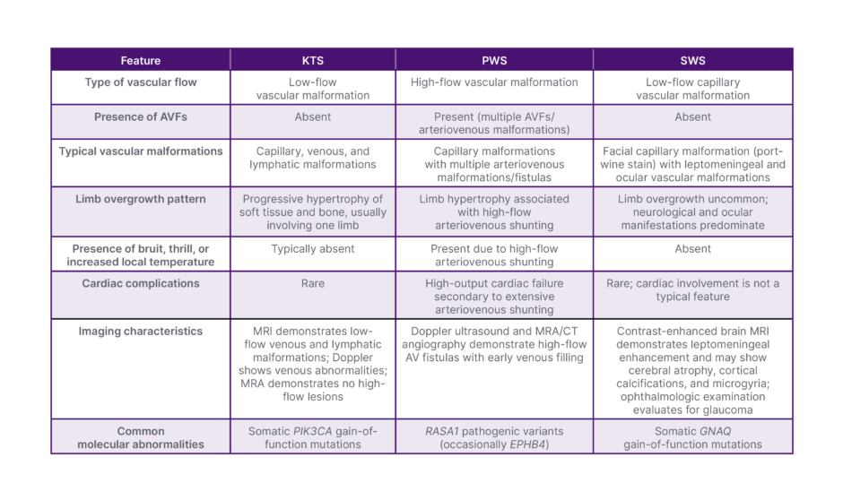
Table 1: Comparison table of KTS, PWS, and SWS based on vascular flow, vascular anomalies, clinical features, imaging findings, and molecular genetics.
AVF: arteriovenous fistulas; KTS: Klippel–Trenaunay syndrome; MRA: magnetic resonance angiography; PWS: Parkes Weber syndrome; SWS: Sturge–Weber syndrome.
The differential diagnoses considered in this case included KTS with/without overlapping SWS and PWS.
Treatment of KTS follows a stepwise approach. Conservative therapy is always the first line, maintained for at least 3–6 months, and includes compression garments, leg elevation, assisted ambulation, lymphatic drainage, wound care, and medications such as aspirin, antibiotics, and anticoagulation as indicated. If conservative measures fail, superficial venous interventions are pursued first, including sclerotherapy, endovenous laser ablation, high ligation and stripping, stab phlebectomy, or radiofrequency ablation, with careful confirmation of deep venous patency before treating the marginal vein. Incompetent perforators may be addressed with coil embolization. For persistent deep system pathology, deep venous interventions are reserved, including iliofemoral venous stenting guided by intravascular ultrasound, popliteal vein release surgery for entrapment, and transcommissural valvuloplasty for refractory deep venous reflux with non-healing ulcers. Perioperative deep vein thrombosis prophylaxis is applied throughout surgical management.
Pharmacological management in KTS is an emerging area, particularly with the development of targeted therapies acting on the PI3K/AKT/mTOR signaling pathway. Sirolimus, an mTOR inhibitor, has shown partial clinical responses in a substantial proportion of patients, with improvements in pain, swelling, and overall vascular lesion burden. However, its use may be limited by adverse effects such as mucositis, fatigue, and rash.2 More recently, newer targeted agents such as miransertib, an AKT inhibitor, and alpelisib, a selective PI3Kα inhibitor, have demonstrated promising outcomes in patients with PROS-related conditions. These therapies have been associated with lesion stabilization as well as improvements in pain, mobility, and chronic bleeding, with alpelisib showing particularly encouraging clinical and radiological responses in early studies.2
Despite these advances, none of these agents are currently formally approved for KTS, and their use remains largely off-label. Careful patient selection and specialist supervision are essential due to potential adverse effects and the need for long-term monitoring. At present, pharmacological therapy serves as an adjunct to conventional management, with further research required to establish its long-term safety and efficacy.
CONCLUSION
This case highlights an unusual dermatological manifestation of KTS, with the presence of hemangiomas, which have not been widely reported in association with this condition. This finding expands the clinical spectrum of cutaneous manifestations within the PROS.
Furthermore, this case underscores the importance of careful clinical assessment supported by radiological imaging in establishing the diagnosis. The initial clinical impression in the authors’ patient suggested a possible arteriovenous malformation; however, imaging findings were instrumental in differentiating KTS from other overlapping vascular disorders including SWS and PWS, which happens to be a high flow arteriovenous malformation syndrome. This approach is particularly relevant in resource-limited settings, where access to advanced molecular diagnostics may be restricted, and diagnosis often relies on a combination of clinical and radiological features.
INFORMED CONSENT
The patient’s family initially expressed anxiety regarding progressive swellings and bleeding skin lesions. After receiving a definitive diagnosis and counseling, they felt reassured and consented to publication to raise awareness about rare vascular disorders. Written informed consent was obtained from the patient’s legal guardians for publication of this case report and accompanying images.