Authors: Kiyan Heybati,1 *Harish Ramakrishna2
1. Department of Internal Medicine, Mayo Clinic, Rochester, Minnesota, USA
2. Department of Anesthesiology and Perioperative Medicine,
Mayo Clinic, Rochester, Minnesota, USA
*Correspondence to [email protected]
Disclosure: The authors have declared no conflicts of interest.
Keywords: Myocardial infarction (MI), myocardial ischaemia, troponin, universal definition
Citation: EMJ Cardiol. 2026;14[1]:22-26. https://doi.org/10.33590/emjcardiol/FV4990B5

ACUTE myocardial infarction (MI) reflects acute myocardial injury, occurring in a setting of myocardial ischaemia, supported by symptoms, ischaemic ECG changes, new loss of viable myocardium on imaging, and/or coronary thrombus.1 The Fifth Universal Definition of Myocardial Infarction (UDMI), developed jointly by the four major cardiac societies: the European Society of Cardiology (ESC), the American College of Cardiology (ACC), the American Heart Association (AHA), and the World Heart Federation (WHF), published 8 years from the last update,1 introduces a revised clinical classification of MI based on pathophysiology, clinical assessment, and objective diagnostic criteria. In this article, the authors review key changes, the underlying rationales, and implications for the practising clinician.
WHAT HAS CHANGED
One of the key changes includes the replacement of the familiar Types 1–5 numerical scheme with a three-part clinical classification (Table 1).2 Notably, as the sensitivity of troponin assays continued to improve, the 2007 definition3 outlined a classification system with five subcategories: Type 1 (atherothrombotic), Type 2 (supply–demand mismatch), Type 3 (cardiac death before biomarkers could be drawn), Type 4 (percutaneous coronary intervention-related, with subtypes for stent thrombosis and restenosis), and Type 5 (coronary artery bypass graft-related). More recently, the fourth iteration outlined a distinction between myocardial injury and myocardial infarction, highlighting that an elevated troponin without other findings suggestive of ischaemia should instead be thought of as injury rather than infarction, which may be acute or chronic.1
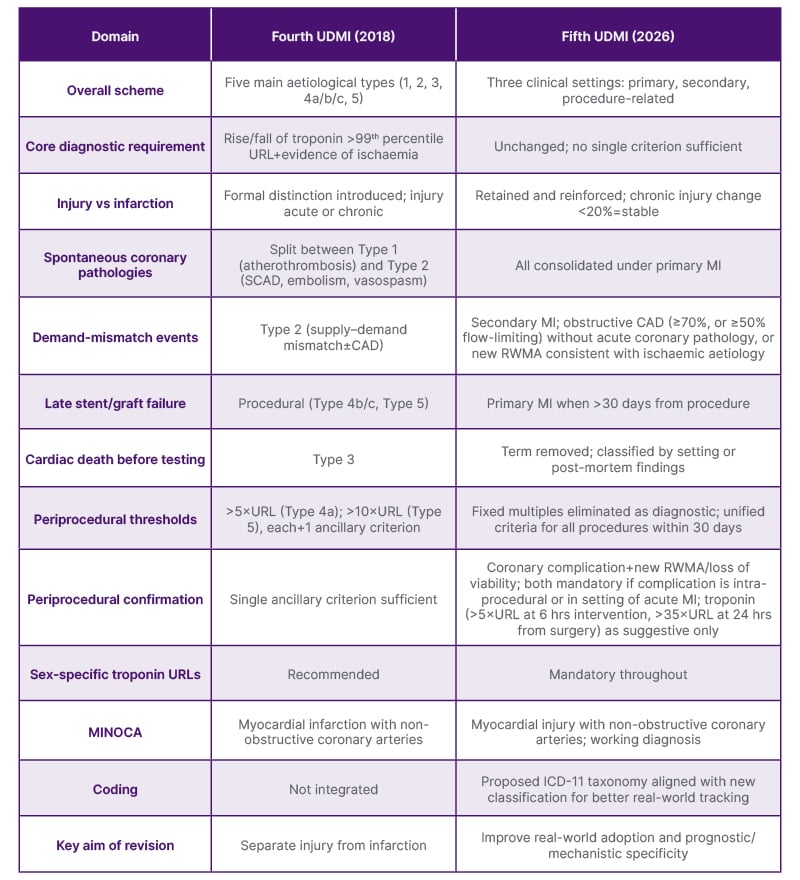
Table 1: Select differences between the Fourth and Fifth Universal Definition of Myocardial Infarction.
CAD: coronary artery disease; ICD: International Classification of Diseases; MI: myocardial infarction; MINOCA: myocardial infarction with non-obstructive coronary arteries; RWMA: regional wall motion abnormalities; SCAD: spontaneous coronary artery dissection; UDMI: Universal Definition of Myocardial Infarction; URL: upper reference limit.
The fifth UDMI comes after studies showed that the earlier classification was inconsistently applied at the bedside. In a two-nation cohort of more than 50,000 patients, a clinical diagnosis of MI was recorded in the majority of adjudicated Type 1 events (79% in Scotland, 87% in Sweden) but in only a minority of adjudicated Type 2 events (17% and 23%, respectively). Notably, patients with an adjudicated Type 1 MI who went unrecognised were more often women and older, yet faced a similar or higher risk of subsequent MI or cardiovascular death.4,5 In addition to this, periprocedural definitions from the fourth UDMI have also been shown to much more frequently classify patients as having a periprocedural MI when compared to the Academic Research Consortium-2 (ARC-2) and Society for Cardiovascular Angiography and Interventions (SCAI) criteria.6,7 Events classified using ARC-2 and SCAI were subsequently shown to hold better prognostic value for cardiac mortality within 1 year versus the fourth universal definition.7 Lindahl and Mills have also noted that the classification scheme had grown to be overly complex, with the potential for adverse outcomes in the event of diagnostic uncertainty.8 Therefore, to better reflect the underlying pathophysiology and decrease the ambiguity often encountered in practice, primary, secondary, and procedure-related definitions have now been formally proposed.2
Primary MI (previously Type 1) covers spontaneous events due to an acute coronary pathology, including aetiologies such as atherothrombosis, spontaneous coronary artery dissection, embolism, and vasospasm, along with restenosis, stent thrombosis, and graft failure occurring more than 30 days after revascularisation (i.e., not regarded as a procedural complication if onset beyond 30 days). Additionally, two further forms of primary MI include undetermined aetiology following angiography or unknown aetiology if no coronary imaging is performed (i.e., based on biomarkers, electrocardiography, and clinical criteria). A key priority is ensuring appropriate sensitivity in diagnosis, with an emphasis on coronary angiography and/or cardiac imaging to further establish the diagnosis and underlying pathology.
Secondary MI (previously Type 2) reflects supply–demand mismatch precipitated by an alternative acute condition as a consequence of underlying coronary artery disease but without evidence of an acute coronary pathology. Notably, the authors highlight that even in acute illnesses such as sepsis or haemorrhage, primary MI should be considered as a differential diagnosis given their tendency to trigger an atherothrombotic event.2 If primary MI is considered a possibility and/or there is evidence of persistent myocardial ischaemia, coronary angiography is indicated with reclassification if an acute coronary pathology is found; however, this may not be feasible in higher-risk patients and requires clinical judgement. Accordingly, definitive confirmation of secondary MI requires either obstructive coronary artery disease (defined as ≥70% stenosis, or ≥50% that is flow-limiting on physiological testing) without an acute coronary pathology or a new regional wall motion abnormality consistent with an ischaemic aetiology. This classification emphasises specificity, supported by a prior study which revealed that such definition would help reduce the diagnosis of MI in acute illness and instead help identify those in the highest risk category for more timely management.9
Procedure-related MI (previously Type 4 and 5) is considered if a patient is within 30 days of the following procedures: coronary angiography with or without intervention, structural cardiac intervention, or catheter ablation, and any open or minimally invasive cardiac surgical procedure, including coronary artery bypass graft, valve replacement, or other procedures for structural heart conditions.2 Rather than primarily relying on troponin thresholds, definitive diagnosis instead requires angiographic evidence of a coronary complication and/or imaging evidence of a new regional wall motion abnormality. Both of these are required for diagnosis if a complication arises during the procedure, or the procedure is performed in the setting of an acute MI. Given that patients may have elevated cardiac biomarkers following intervention, while some studies10 have sought to outline thresholds for diagnosing clinically relevant periprocedural myocardial injury, the sensitivity and negative predictive value of such cut-offs remain relatively understudied.
Moreover, other notable changes include the removal of Type 3 MI, with cardiac death before testing now being classified by setting or post-mortem findings into one of the three clinical categories outlined above. MINOCA has also been redefined from ‘myocardial infarction’ to ‘myocardial injury with non-obstructive coronary arteries’. This change is meant to highlight MINOCA as a working diagnosis requiring further evaluation rather than an endpoint.11-13 Specifically, prior studies have suggested that a notable proportion of patients are ultimately found to have a non-coronary cardiac or non-cardiac cause. Lastly, yet another notable change includes proposed ICD-11 codes for each MI aetiology/setting for better epidemiological tracking and real-world application for subsequent management strategies such as risk-factor modification and rehabilitation.2
WHAT HAS STAYED THE SAME
Overall, the foundational concepts have remained intact, with acute myocardial injury still defined as a rise and/or fall in cardiac troponin with at least one value above the 99th percentile upper reference limit, and infarction requiring that injury with additional evidence of ischaemia. The delineation between injury and infarction is also reinforced, with chronic injury retaining its own criteria. Additionally, while previously recommended, sex-specific 99th percentile upper range limits for biomarkers are now mandatory. This is meant to avoid systematic bias and the under-recognition of myocardial injury and infarction in women, which has been demonstrated in prior studies.5
IMPLICATIONS FOR PRACTICE
For the practising clinician, the fifth iteration proposes several changes to decrease diagnostic ambiguity and, in turn, leads to a better understanding of underlying pathology and subsequent management. This is primarily achieved through a new classification system focusing on primary, secondary, and procedure-related MI. Additionally, definitive diagnostic criteria for each further emphasise imaging as a part of objective confirmation, primarily through the detection of new regional wall motion abnormalities. This is especially pertinent in the setting of procedure-related MI where troponin elevation cut-offs, while suggestive, are meant to trigger further diagnostics rather than to confirm the diagnosis. At the same time, in resource-limited settings where imaging may not always be readily available, a pragmatic framework is outlined based on clinical presentation, ECG, and biomarkers. Moreover, in response to findings that women were less likely to be appropriately diagnosed with MI, sex-specific troponin thresholds are now required. Acknowledging that further investigation in this area is necessary, a set of ICD-11 codes is proposed to enable further epidemiological tracking as well as facilitating appropriate management including risk factor modification and rehabilitation.