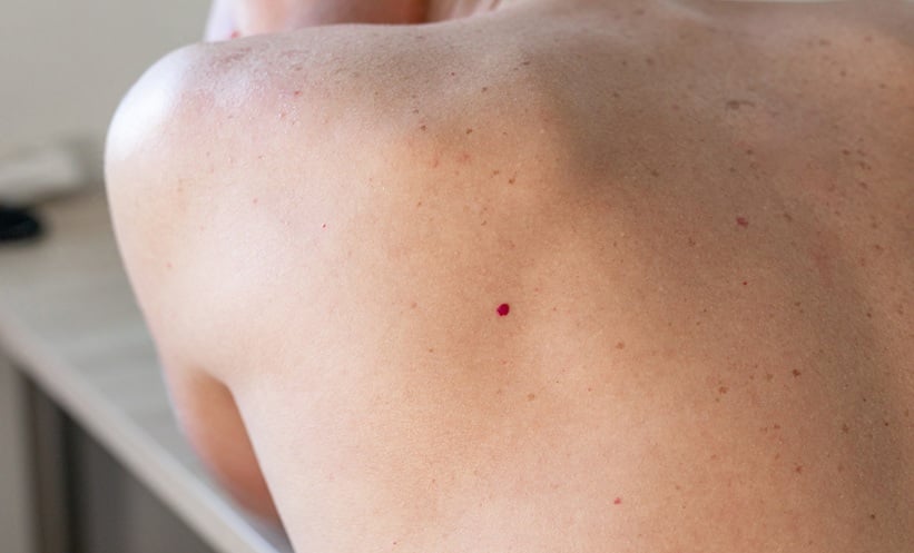
CHERRY angiomas were significantly more prevalent and developed at a younger age in individuals with neurofibromatosis type 1 (NF1), with genetic analyses suggesting these lesions may represent a previously unrecognised vascular manifestation of the condition.
The prospective cross-sectional study included 259 participants aged 15 years or older, comprising 102 individuals with confirmed NF1 and 157 controls. The epidemiological analysis was conducted at a French national referral centre for neurofibromatoses between October 2020 and March 2021, followed by histopathological, genomic, and cell-specific molecular analyses.
Cherry angiomas were identified in 48% of individuals with NF1 compared with 18% of controls. The association remained after adjustment for age and sex and in propensity score-matched analyses.
The odds of having a cherry angioma were also higher among individuals with NF1 (odds ratio: 4.26; 95% CI: 2.44–7.56).
NF1 Variants Identified in Lesions
The researchers investigated the biological basis of the association by analysing cherry angiomas from individuals with NF1 and healthy volunteers.
Somatic NF1 loss-of-function second-hit variants were identified in 26 of 39 NF1-associated cherry angiomas (67%), while no such variants were identified in control lesions. This indicated biallelic NF1 inactivation within the NF1-associated lesions.
Genomic profiling also identified co-occurring activating variants, with GNAQ among the most frequently observed. Cell-specific sequencing indicated that NF1 second hits arose predominantly in endothelial cells and telocytes, with higher variant allele frequencies observed in endothelial cells.
Potential Vascular Manifestation of NF1
Immunofluorescence analysis demonstrated increased phosphoextracellular signal-regulated kinase signalling in the vascular cell populations carrying NF1 alterations, providing further evidence of pathway activation within the lesions.
The researchers suggested that the findings extend understanding of NF1-associated manifestations beyond neural crest-derived tissues and support cherry angiomas as a potential model for NF1-related vasculopathy.
Although the study was cross-sectional, the combination of epidemiological and mechanistic findings suggests that the increased occurrence of cherry angiomas in individuals with NF1 may have a biological basis rather than representing an incidental clinical observation.
The findings could broaden the recognised spectrum of NF1-associated neoplasms and provide a model for investigating vascular disease associated with NF1 loss.
Reference
Fertitta L et al. Cherry angiomas in individuals with neurofibromatosis type. JAMA Dermayol. 2026;DOI:10.1001/jamadermatol.2026.2933.
Featured image: Olya on Adobe Stock