Abstract
Early mammalian embryogenesis is driven by a tightly regulated transition from maternally inherited transcripts to zygotically transcribed RNA, a process that requires precise coordination of RNA stability, translation, and selective clearance of maternal transcripts. Emerging evidence places epitranscriptomic regulation at the centre of these mechanisms. This review summarises current knowledge of key RNA modifications, their regulatory machineries, and their functional roles during early mammalian development. Specifically, emphasis is placed on internal base modifications, including N6-methyladenosine, N4-acetylcytidine, and 5-methylcytidine, as well as RNA editing (A-to-I and C-to-U), RNA capping, and cap-proximal methylations. Many RNA modifications are dynamic and reversible, modulating RNA structure, stability, and translational efficiency in a stage-specific manner. Genetic and functional studies demonstrate that these modifications, deposited, decoded, and removed by dedicated writer, reader, and eraser proteins, are essential for zygotic genome activation, preimplantation development, mitochondrial function, cellular fitness, and early lineage specification. Cap formation and cap-adjacent methylations add an additional regulatory layer by coupling mRNA export and translation to maternal RNA decay and the onset of zygotic gene transcription. Furthermore, dynamic RNA editing expands transcriptomic and proteomic diversity, enabling context-dependent gene regulation without altering the underlying DNA sequence. Collectively, these findings establish RNA modifications and RNA processing as key determinants of developmental competence. In the future, modification-specific targets, context-dependent functions, and the mechanisms of epitranscriptomic crosstalk still need to be elucidated, presenting new avenues for the diagnostic and therapeutic targeting of RNA modifications in reproductive medicine.
Key Points
1. Early mammalian development depends on accurate maternal RNA clearance and zygotic activation; disrupted RNA modification or decay compromises developmental competence and contributes to infertility and recurrent miscarriage.2. This narrative review integrates systematically screened publications on key RNA modifica-tions, capping, and RNA editing, outlining their writer–reader–eraser machineries and stage-specific functions in early mammalian embryogenesis.
3. Epitranscriptomic regulation of early embryogenesis is an emerging field, with the potential to improve diagnosis of RNA-driven infertility and pregnancy loss and guide future reproduc-tive therapies.
INTRODUCTION
In mammals, early embryonic development begins at fertilisation, when the oocyte and sperm fuse to form the one-cell zygote. The one-cell embryo subsequently undergoes a series of cleavage divisions, during which development is initially driven largely by maternally inherited transcripts and proteins accumulated during oogenesis.1 Later, a minor wave of zygotic genome activation (ZGA) occurs at the late one-cell stage. This minor ZGA is characterised by widespread RNA polymerase II recruitment and pervasive, often intergenic transcription, frequently initiated outside canonical transcription start sites, producing short, frequently unspliced transcripts. Despite the necessity of the minor ZGA for further embryo development, this stage is still characterised by an abundance of maternal transcripts. As development proceeds, control shifts from maternal to embryonic gene products, a process referred to as maternal-to-zygotic transition (MZT). MZT consists of a series of molecular events, beginning with the degradation of subsets of maternally deposited RNAs. This maternal RNA clearance occurs in two waves. The first, M-decay, which represents maternal factor-regulated decay, initiates after germinal vesicle breakdown and is sustained until the metaphase II stage before fertilisation.2 The second wave, Z-decay, depends on the expression of specific early zygotic factors for degrading maternal mRNA. Z-decay is coupled to the major ZGA, which features more canonical transcription initiating at defined transcription start sites and regulated by splicing and elongation factors. The major ZGA drives active transcription of hundreds of genes required for Z-decay (Figure 1A).2
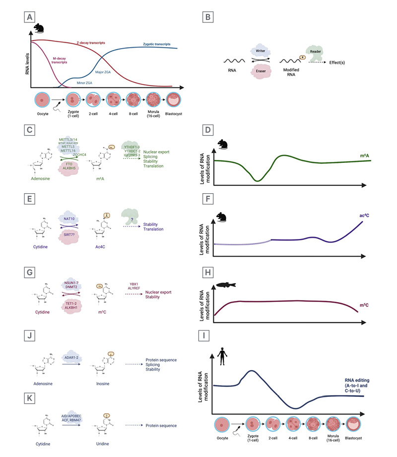
Figure 1: Overview of RNA modifications discussed in this paper.
A) Schematic overview of oocyte and embryo RNA levels in mice, indicating important events such as M- and Z-decay and major and minor ZGA. B) Schematic representation of the function of writer proteins (blue), eraser proteins (red) and reader proteins (green). C) Chemical structure of m6A, along with writers, erasers, readers, and established functions of the modification. D) Schematic representation of m6A modification levels through embryonic development in mice.14 E) Chemical structure of ac4C along with writers, and potential eraser, and established
functions of the modification. F) Schematic representation of ac4C modification levels through embryonic development in mice. Dotted line represents levels implied from non-quantified immunofluorescence images.38
G) Chemical structure of m5C, along with writers and established functions of the modification. H) Schematic representation of m5C modification levels through embryonic development in zebrafish.42 I) Schematic representation of editing events through embryonic development in humans.50 J) Chemical reaction of A-to-I editing, writers, and established functions of the modification. K) Chemical reaction of C-to-U editing, writers and established functions of the modification.
Created in BioRender.
Ac⁴C: N⁴-acetylcytidine; ACF: APOBEC-1 complementation factor; ADAR1-2: adenosine deaminase acting on RNA 1 and 2; AID: activation-induced cytidine deaminase; ALKBH1: AlkB homolog 1; ALKBH5: AlkB homolog 5; ALYREF: Aly/REF export factor; APOBEC: apolipoprotein B mRNA editing enzyme, catalytic polypeptide-like; DNMT2: DNA methyltransferase 2; FTO: fat mass and obesity-associated protein; IGF2BP1-3: insulin-like growth factor 2 mRNA-binding proteins 1–3; m⁵C: 5-methylcytidine; m⁶A: N⁶-methyladenosine; METTL3: methyltransferase-like 3; METTL16: methyltransferase-like 16; NAT10: N-acetyltransferase 10; NSUN1-7: NOP2/Sun RNA methyltransferases 1–7; RBM47: RNA-binding motif protein 47; SIRT7: sirtuin 7; TET1-2: ten-eleven translocation methylcytosine dioxygenases 1 and 2; WTAP: Wilms tumour 1-associated protein; YBX1: Y-box-binding protein 1; YTHDC1-2: YTH domain-containing proteins 1 and 2; YTHDF1-3: YTH N⁶-methyladenosine RNA-binding proteins 1–3; ZCCHC4: zinc finger CCHC-type containing 4; ZGA: zygotic genome activation.
The timing of these events varies between species. The major ZGA occurs in the two-cell stage in mice, and Z-decay follows, lasting from two-cell to four-cell stage. In humans, ZGA is broader and less sharply defined, occurring around the four-cell to eight-cell stage, and the Z-decay spans approximately from the four-cell to morula stage.2 However, both species alike undergo at least two waves of maternal RNA-clearance.3,4
In mice and humans, germ cells initiate meiosis during embryonic development and arrest at the germinal vesicle stage until puberty. Upon the pre-ovulatory hormone surge, these fully grown oocytes resume meiosis, transitioning from the germinal vesicle stage to metaphase II arrested stage. Maternal RNAs accumulate during oocyte growth and maturation and remain translationally dormant until meiotic resumption.5,6 Upon fertilisation, maternal RNAs can be actively translated or degraded within a few hours to days. Substantial advances have deepened our understanding of how RNA stability is regulated in mammalian oocytes and zygotes. MZT transcript fate is governed by interconnected mechanisms involving sequence elements, RNA-binding proteins, poly(A) tail dynamics, and subcellular localisation. Beyond these established pathways, accumulating evidence highlights covalent RNA modifications as an additional layer of post-transcriptional control.
This expanding field, known as epitranscriptomics, investigates how chemical marks on RNA interact with regulatory networks to fine-tune gene expression. ‘Writer’ enzymes deposit chemical modifications, either co-transcriptionally or post-transcriptionally, whereas ‘eraser’ enzymes remove RNA modifications.7 Epitranscriptomic marks influence RNA processes such as stability, translation, and splicing through interactions with RNA-binding ‘reader’ proteins (Figure 1B). RNA can be modified in several ways, including the addition of chemical groups, editing of RNA sequences, and capping of RNA ends.7
Although RNA modifications have been known since the 1970s, only the recent development of sensitive detection techniques has revealed their widespread occurrence, dynamic potential, and biological significance. More than 170 distinct RNA modifications have been identified, yet the specific role of most of these marks remains unclear. RNA modifications hold considerable promise as diagnostic biomarkers and therapeutic targets, with the potential to improve fertility and outcomes of assisted reproductive technologies. In this context, a comprehensive literature review was conducted to evaluate current evidence of the roles of epitranscriptomics in preimplantation development and to advance our understanding of early mammalian embryogenesis.
METHODS
During the preparation of this review, the scientific literature was searched using PubMed/Medline between January–March 2026. Initially, a broad search was conducted with the following search term: (“epitranscriptomic*” OR “RNA modification*”) AND (“gametogenesis*” OR “embryo*” OR “embryogenesis*” OR “embryo development*” OR “mammalian development*”). The resulting primary research papers and review articles were screened based on titles and abstracts as presented in Figure 2A. For review articles specifically, a stringent inclusion criterion based on publication date was applied to ensure that only the most recent summary works were included. The selected reviews and primary papers were then skimmed for relevance and used to provide background information and to determine which RNA modifications to cover, focusing on those that were the most well established and thoroughly investigated.
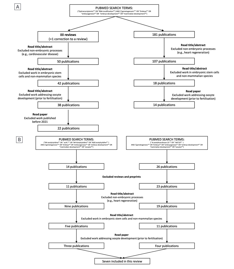
Figure 2: Schematic example of workflow.
*Asterisks are included in the search terms to find variations of a phrase or word. E.g., “Embryo*” will find “Embryo,” “Embryos,” “Embryonic,” and “Embryology.”
A) The workflow used for an initial broad search used for compiling initial background information. B) The simplest
example of the workflow used for later specific searches for each RNA modification.
Figure 2B illustrates the workflow applied to individual searches conducted for each RNA modification, as well as for known reader, writer, and eraser proteins (all search term results are listed in Supplementary Table 1). Identified articles were similarly screened based on titles and abstracts. Inclusion criteria were English language, primary research, and meta-analyses published in peer-reviewed journals. Preprints (e.g., from bioRxiv) were also screened for relevance. Exclusion criteria included review articles and studies that did not address epitranscriptomics or the selected RNA modifications in early mammalian development. Studies conducted in non-mammalian species or in embryonic stem cells were reviewed but excluded unless no relevant studies in mammals were available. Papers passing this initial screening were skimmed and excluded if, on closer reading, they did not address the function of the relevant RNA modifications or associated proteins, or if they otherwise met any of the exclusion criteria. For all studies meeting the inclusion criteria, relevant information was manually extracted. The authors reviewed all papers included in the final manuscript.
RESULTS
Chemical RNA Modifications in Embryogenesis
Chemical modifications that decorate RNA are diverse and include, for example, methylation and acetylation. These modifications can influence RNA structure, base pairing, and interactions with other molecules, thereby modulating RNA stability and function.7
N6-methyladenosine
N6-methyladenosine (m6A) is the most abundant internal modification on mammalian mRNA.7 It is installed by a multicomponent methyltransferase complex composed of two subcomplexes of the catalytic subunit m6A-METTL complex (MAC) and the regulatory subunit m6A-METTL-associated complex (MACOM). The catalytic core, MAC, consists of a METTL3–METTL14 heterodimer that transfers a methyl group from S-adenosyl-L-methionine to the N6 position of adenosine residues.8 MACOM confers substrate specificity and includes components such as WTAP and KIAA1429.7 Additional m6A methyltransferases include METTL16, which modifies structured RNAs containing hairpin motifs;9 METTL5, which catalyses m6A modification of rRNA;10 and ZCCHC4, which methylates 28S rRNA.11,12 m6A marks are recognised by specific RNA-binding ‘reader’ proteins, including YTH domain-containing proteins (YTHDF1–3 and YTHDC1–2) and insulin-like growth factor 2 mRNA-binding proteins (IGF2BP1–3).7 m6A levels are dynamically regulated by the demethylases fat mass and obesity-associated protein (FTO) and ALKB homolog 5 (ALKBH5), which catalyse oxidative removal of the methyl group (Figure 1C).7,13
The picogram-scale m6A RNA immunoprecipitation and sequencing method (pico-MeRIP-seq) was recently developed, enabling the first systematic characterisation of the human m6A landscape during early embryogenesis.14,15 In human embryos, the number and percentage of m6A modified genes steadily decrease until fertilisation, reaching a low point at the one-cell stage, followed by an increase from the two-cell stage and a plateau throughout the eight-cell to blastocyst stage.14 A recent study, using crosslinked-antibody RNA immunoprecipitation and sequencing, has suggested that m6A levels increase through the pre-fertilisation stages, peaking at the zygote stage, after which it drops, before increasing again from four-cell to blastocyst stage.16
In mice, the m6A landscape follows a similar pattern: the number of m6A modified genes declines until fertilisation, before increasing again as the embryo progresses through the first cell divisions (Figure 1D).17,18 Single-cell sequencing suggests that m6A levels may rise after meiosis II and remain relatively constant from the zygote stage onward.19 In contrast, mass-spectrometry based quantification indicates that m6A levels drop after the germinal vesicle stage and stay low throughout the first two cell divisions,18 while others have recently reported relatively constant m6A levels throughout embryogenesis.16
However, the percentage of the specific m6A modified genes varies between human and mouse, likely reflecting evolutionary divergence.14 m6A can be either maternally inherited or deposited de novo after fertilisation. Maternally inherited m6A modifications serve dual functions: they promote the decay of a subset of transcripts while stabilising others and licensing them for translation upon fertilisation.20
The Role of m6A Writers
The importance of m6A writers in embryonic development has been demonstrated by depleting core components. In mice, Mettl3 knockout (KO) abolished m6A-signals19 and resulted in developmental arrest at the four-cell stage.21 Pharmacological inhibition of METTL3 with STM2457 or cycloleucine similarly reduced m6A levels and delayed development at the four-cell stage, markedly decreasing blastocyst formation.18,22 Mechanistically, METTL3 preferentially mediates de novo establishment of m6A on ZGA transcripts without influencing m6A on maternal transcripts.18 METTL3 primarily methylates transcripts with transient expression, which are downregulated after the two-cell stage,18 and its inhibition upregulates these genes at the four-cell and morula stages due to the lack of m6A promoting mRNA-decay.18 Prior to fertilisation, m6A stabilises transcripts, and Mettl3 knockdown (KD) reduces m6A levels, resulting in downregulation of numerous genes in germinal vesicles,21 which in turn leads to decreased transcription-engaged RNA polymerase and globally reduced transcription.23 METTL3 and global m6A levels were significantly decreased in placentas from patients with fetalgrowth restriction.24
Other m6A writers also play crucial roles in embryogenesis. Mettl16 KO mice fail to develop beyond the blastocyst stage, exhibiting thousands of differentially expressed genes, which suggests a role in later stages of early embryonic development.9 In contrast, METTL5 depletion can enhance certain developmental outcomes. siRNA-mediated Mettl5 KD in somatic cell nuclear transfer (SCNT) embryos reduced m6A peaks but increased cell numbers and improved blastocyst formation.25 This effect is proposed to result from METTL5-mediated rRNA methylation, which alters ribosomal translation of histone methyltransferases and thereby modulates two-cell stage gene expression.25
MACOM components likewise influence m6A and embryogenesis. Kiaa1429 KO mice experience lower global m6A-levels, with a pronounced effect on Z-decay genes18 and WTAP KD reduced m6A levels and blastocyst formation rate in porcine embryos, accompanied by decreased pluripotency markers.26
The Role of m6A Readers
m6A readers, which recognise the modification and mediate cellular responses, are also essential for early embryonic development. Female mice lacking Ythdf2 are infertile, as loss of Ythdf2 causes an abnormal two-cell stage, preventing further development.18,27 Consequently, Ythdf2-depleted embryos exhibit impaired RNA degradation,27 resulting in increased expression of transcripts normally targeted during Z-decay.18 Similarly, in goat embryos, siRNA-mediated KD of YTHDF2 blocks blastocyst formation, with embryos arresting at the four- and eight-cell stages due to defective maternal mRNA clearance and ZGA.28
Heterozygous Ythdc1 mutant mice fail to produce homozygous Ythdc1 KO offspring, with embryos being reabsorbed from embryonic day E8.5.29 Igf2bp1 KD reduces m6A levels in parthenogenetic embryos from mice,30 and is associated with lower blastocyst formation rate in both parthenogenetic mouse embryos and porcine embryos.30,31 Meanwhile, maternal depletion of Igf2bp2 in mice leads to severe defects in blastocyst formation, with embryos arresting at the two-cell stage and exhibiting reduced RNA and protein synthesis.32
The Role of m6A Erasers
The two established m6A erasers are FTO and ALKBH5. Studies on FTO in embryogenesis have yielded conflicting results. Wei et al.33 reported that IVF rates are more than halved when wild-type oocytes are fertilised with Fto mutant sperm compared to controls.33 However, when oocytes were fertilised by intracytoplasmic sperm injection using Fto mutant sperm, the resulting embryos exhibited only slightly impaired progression to the blastocyst stage but failed to implant.33 This effect of FTO has been attributed to FTO-mediated m6A removal on LINE1 RNA, and that in turn shapes the global chromatin landscape.33 Contrarily, Kim et al.34 has found that deletion of the Ft-locus (which contains Fto along with five other genes), did not affect early embryonic development, but did result in a lower primordial germ cell proliferation.34 By contrast, ALKBH5 appears to be essential for fertility. Alkbh5 mutant mice are infertile due to meiotic defects that arrest oocyte development35 and metaphase-stage spermatocytes.13 Additionally, lower expression levels were detected in villous tissue of patients with reoccurring miscarriages compared to healthy controls.36
N4-acetylation of cytosine
A much less studied RNA modification is N4-acetylation of cytosine (ac4C), for which only one writer, N-acetyltransferase10 (NAT10), has been identified, and no readers or erasers are known to date. ac4C has been shown to enhance the stability and translation of RNAs, thereby modulating gene expression (Figure 1E).37
ac4C levels in early embryonic development have been determined by immunofluorescent staining, showing stable levels from two-cell to morula stage, with a drastic increase at the blastocyst stage (Figure 1F).38
Since NAT10 is the only known ac4C writer, and its depletion markedly reduces ac4C levels in oocytes,39 most functional studies of this acetylation in early embryos have focused on manipulating NAT10 level. Both siNAT10 and dsNAT10 treatment in mouse embryos has been shown to decrease blastocyst formation, arresting embryos at the morula stage,40,41 and causing a cavity collapse and degeneration.38 Furthermore, Nat10 KD in morula resulted in significantly lower expression of the pluripotencyfactor Nanog.40
5-methylcytosine
5-methylcytosine (m5C) is generated when cytosine is methylated at the 5th carbon by RNA m5C methyltransferases, primarily the NOL1/NOP2/SUN (NSUN) family and the DNA cytosine-5 methyltransferase 2 (DNMT2). To date, two m5C readers have been identified: Y-box binding protein 1 (YBX1) and Aly/REF export factor (ALYREF; Figure 1G).8
The m5C landscape has been mapped across embryonic stages in several species, revealing a conserved pattern of high m5C levels at the one-cell stage, followed by a sharp decline after the MZT (Figure 1H).42
In mice, Nsun2 KO impairs ovarian development, and siRNA-mediated KD of Nsun2 in oocytes blocks development at the two-cell stage, leading to reduced blastocyst formation.43 NSUN3 has also been shown to be important for embryonic development, albeit at later stages, as Nsun3 KO mice die between embryonic day E10.5 and E12.5.44 NSUN4 is a mitochondrial m5C-methyltransferase that methylates rRNA. Its deletion has been shown to cause embryonic lethality in mice, with embryos showing retarded growth with no clearly discernible anatomical structures at E8.5.45 Nsun5 KO mice produce fewer offspring, with compromised embryogenesis from the one-cell to blastocyst stages, an effect attributed to loss of NSUN5-mediated m5C targets.46 Consistently, Nsun5 KD in mice reduced morula cell number and blastocyst formation, impaired differentiation and trophectoderm development.47
YBX1 KD reduces developmental rates, with decreased blastocyst formation and cell numbers, and impairs maternal mRNA degradation and ZGA in both goat and porcine embryos.31,48 Another m5C reader, ALYREF, is also important in early development: embryonic KD of Alyref impairs blastocyst formation due to insufficient Nanog expression, likely by destabilising the mRNA.49
RNA Editing in Embryogenesis
RNA editing is a co- or post-transcriptional modification by which cells alter the nucleotide sequence of RNA. During human embryonic development, A-to-I and C-to-U edits are the most common, accounting for approximately 86% of all editing events.50 RNA editing exhibits distinct temporal patterns throughout development, with specific sites differentially edited at different stages: some sites are highly edited only at the zygote stage, others remain highly edited until the four-cell stage, and some are edited predominantly at the eight-cell stage (Figure 1I).50 These patterns suggest that RNA editing is stage-specific and may play a role in regulating RNA splicing and gene expression.
Adenosine to inosine
A-to-I editing, one of the most prominent RNA-editing events, involves the conversion of adenosine to inosine in double-stranded RNA. In oocytes, this editing occurs more frequently in protein-coding sequences at the codon wobble position,51 potentially altering protein sequences because translating ribosomes read inosine as guanosine. A-to-I editing is catalysed by adenosine deaminases acting on RNA (ADAR) 1 and 2 (Figure 1J).7
Recent analyses of RNA-seq datasets have identified recurrent A-to-I edits throughout human embryogenesis. Two independent studies reported that A-to-I-edited transcripts are present at all early embryonic stages, but the levels of inosine-containing transcripts decline after the four-cell stage.52,53 Ding et al.52 further observed that A-to-I-edited transcripts tend to show reduced expression in later embryonic stages, proposing that this reduction is partly mediated by microRNA-induced degradation, as A-to-I-edited mRNAs gain microRNA binding sites.52 Given that ADAR proteins catalyse A-to-I editing, their expression patterns are expected to reflect editing dynamics. Indeed, ADAR1 expression mirrors A-to-I editing: high from the zygote to four-cell stage, followed by a drop at the eight-cell stage.53 Bovine embryos conceived via IVF display a similar ADAR expression profile, with high levels at the two-cell stage and a decline at the blastocyst stage, whereas this dynamic pattern is absent in cloned embryos from SCNT.54 These observations suggest that proper A-to-I editing may be one reason why SCNT cloning has proven challenging.
In mice, both Adar1 KO animals and mice carrying an editing-deficient ADAR1 mutation (ADAR1E861A) die during embryonic development.55-57 This lethality has been attributed to a failure of RNA editing, which normally introduces destabilising mismatches that prevent dsRNA accumulation. In its absence, endogenous dsRNA builds up, triggering aberrant dsRNA mediated interferon signalling, defective haematopoiesis, and embryonic death around E13.5.56 Consistent with an essential role of ADAR1 in fertility, granulosa cell-specific deletion of Adar1 results in delayed ovulation, impaired oocyte maturation, and infertility in mice.58 Adar2 KO mice are produced at Mendelian ratios from heterozygote intercrosses, but die between P0 and P20 due to defective A-to-I editing of a glutamate receptor mRNA.59
Cytidine to uridine
C-to-U editing is catalysed by members of the activation-induced cytidine deaminase/apolipoprotein B mRNA-editing enzyme catalytic polypeptide-like (AID/APOBEC) family. Mechanistically, C-to-U editing involves the deamination of cytidine to uridine, which can alter the coding sequence of the mRNA (Figure 1K).60 The most well-known example is the editing of APOB mRNA, where a single C-to-U change introduces a premature stop codon, producing the truncated APOB48 protein.60 To date, few studies have investigated the role of C-to-U RNA editing during embryonic development. Those that have, focused primarily on cofactors of APOBEC1, notably apobec-1 complementation factor (ACF) and RNA-binding motif protein 47 (RBM47), which guide editing specificity. RBM47 is required for C-to-U editing, as demonstrated by impaired Apob editing in Rbm47-deficient mice.61 These mice exhibit increased mid-gestation embryonic lethality, and surviving Rbm47-deficient pups are consistently smaller than their wild-type littermates.62 Similarly, ACF is essential for preimplantation development, as Acf-deficient embryos fail to develop past the blastocyst stage.63 Because Apobec1-deficient mice are viable,64 the embryonic lethality observed in Rbm47– and Acf-deficient animals may be independent of APOBEC1-mediated C-to-U editing.
RNA Capping and Cap Proximity Methylations in Embryogenesis
RNA products of RNA polymerase II, including mRNAs, are rapidly capped at the 5′ end to protect them from cellular exonucleases. Capping is a multistep, enzyme-catalysed process initiated by the addition of guanosine to the nascent transcript by RNA-triphosphatase and guanylyltransferase. Subsequently, RNA guanine-7-methyltransferase methylates the cap guanosine at the N7 position, generating the 7-methylguanosine (m7G) cap (Figure 3A-C).7 m7G caps can be removed by decapping enzymes, primarily the mRNA-decapping (DCP)1–DCP2 complex (Figure 3D).65
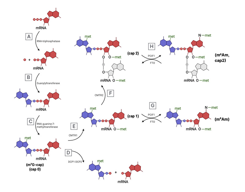
Figure 3: Mechanism of RNA capping, decapping, and cap-proximity methylations.
A) RNA-triphosphatase removes the γ-phosphate from the 5’-ppp-RNA forming 5’-pp-RNA. B) Guanyltransferase couples a GMP, from GTP, to the 5’-pp-RNA molecules. C) RNA guanine-7-methyltransferase transfers a methyl group from SAM to the N7 of the terminal guanine to generate the m7G-cap (cap 0). D) m7G-capped RNA can be decapped by the DCP1–DCP2 complex. The catalytically active DCP2 hydrolyses one of the triphosphate bonds between the RNA-molecule and the m7G-cap. E) Alternatively, the m7G-capped RNA can be methylated near the cap. CMTR1 mediates the 2-O-methylation of the ribose in the most proximal nucleotide to the m7G-cap, forming the cap 1 structure. F) This structure can be further 2-O-methylated by CMTR2, methylating the ribose of the second most proximal nucleotide to the m7G-cap, forming the cap 2 structure. Both RNA with cap 1 G) and cap 2 H) structures can be modified by PCIF1, if the proximal nucleotide is adenosine. PCIF1 methylates the N6 of the adenosine forming m6Am, a reaction that can be reversed by FTO.
Created in BioRender.
CMTR1: cap methyltransferase 1; CMTR2: cap methyltransferase 2; DCP1: mRNA-decapping enzyme 1; DCP2:
mRNA-decapping enzyme 2; FTO: fat mass and obesity-associated protein; GMT: guanine-N7 methyltransferase; m⁶A: N⁶-methyladenosine; m⁶Am: N⁶,2′-O-dimethyladenosine; met: methyl group; PCIF1: phosphorylated CTD-
interacting factor 1.
Cap-proximal methylation refers to methylations occurring near the 5′ cap. Cap methyltransferase (CMTR)1 and CMTR2 catalyse 2′-O-ribose methylation of the first and second nucleotides, respectively (Figure 3E–F). When the first nucleotide is adenosine, the cap-specific adenosine methyltransferase Phosphorylated CTD Interacting Factor 1 (PCIF1) can methylate the N6 position of 2′-O-methyladenosine, generating N6-O-dimethyladenosine (m6Am), a modification that can be removed by FTO (Figure 3G–H).7 While most genes involved in m7G capping are expressed throughout early embryonic development,66 not all have been thoroughly investigated in the context of embryogenesis.
The m7G cap binding complex mainly responsible for the nuclear export of mRNA, is composed of nuclear cap binding protein (NCBP)1 and NCBP2. In mice, lack of Ncbp1 results in developmental arrest in the morula stage and is associated with RNA-retention in the nucleus.67
eIF4F is a cap binding complex facilitating steady translation initiation, and is composed of eIF4G, enabling recognition of the poly-A tail and eIF4E, responsible for m7G cap binding. In mice, maternal eIF4E is carried over from the oocyte, but is rapidly degraded upon fertilisation. By the late two-cell stage, when the embryonic gene expression is assured, eIF4E is crucial for protein synthesis.68 Mechanistic studies have shown that inhibiting maternal eIF4E in the zygote stage prevents the development beyond the two-cell stage.68
eIF4E1B is the germinal cell specific eIF4E isoform.69 Eif4e1b is expressed in oocytes and zygotes, but expression diminishes by the two-cell stage.70 Genetic excision of eif4e1b leads to developmental arrest in the two-cell stage in mice70,71 due to impaired transcription of genes activated during the ZGA.71 Additionally, eIF4E1B binds target mRNA, preventing their degradation in metaphase II-stage oocytes,71 which are then, upon fertilisation, translated with a higher efficiency.70 Since eIF4E1B targeted mRNA include chromatin remodelling factors,70 a loss of eIF4E1B is associated with a failure to reset zygotic chromatin to an open structure, leading to failed activation of zygotic genes.71
In humans, mutations in eIF4E nuclear import factor 1 (eIF4ENIF1), a protein critical for eIF4E nucleocytoplasmic shuttling, are associated with autosomal dominant inherited primary ovarian insufficiency and early menopause.72
DCP1–DCP2 form the catalytic core of the m7G decapping complex. In mice, Dcp1a and Dcp2 mRNA are highly expressed in oocytes and one-cell stage, but expression drops thereafter,73 suggesting these transcripts are maternally expressed and loaded into the oocyte and not expressed during the ZGA. siRNA-mediated KD of both Dcp1a and Dcp2 increases maternal mRNA stability, elevating levels of transcripts usually degraded in the two-cell embryo.73 This, in turn, inhibits the ZGA, as evidenced by lower overall transcription levels and active promoter marks in two-cell embryos.73 Using combined RNA-seq and proteomics analysis, Kong et al.74 confirmed that accumulation of DCP1A coincided with increased degradation of maternal mRNA.74 Increased DCP1A protein is observed in postovulatory aged mouse and human oocytes, and suppressing this rise improves oocyte quality, as evidenced by reduced fragmentation and increased blastocyst formation.75
Cap-modifiers have also been shown to be important in embryonic development. Both CMTR1 and CMTR2 are critical for embryonic development, as KO leads to embryonic lethality in mice.76 Cmtr1 and Cmtr2 KO embryos showed no apparent phenotype until E6.5 but displayed signs of arrested development by E7.5.76 Pcif1 KO mice, displaying a prominent decline in m6Am levels, shows no obvious effect on fertility or early development.77 Recent profiling of the m6Am landscape in both mouse and human embryogenesis has shown that it is a highly dynamic process, with m6Am levels reaching a low point around the time of the ZGA.16 Furthermore, it was proposed that m6Am promotes RNA expression and translational efficiency, potentially to a stronger degree than m6A, and can act synergistically with other modifications.16
CONCLUSION
Over the last decade, numerous studies have demonstrated that early embryogenesis is not governed solely by transcriptional control; it critically depends on a complex epitranscriptomic layer. Loss-of-function studies in animals demonstrate that perturbing individual writers, readers, erasers, or cap/decapping factors often results in preimplantation arrest, abnormal blastocyst formation, or impaired exit from pluripotency. These findings indicate that epitranscriptomic regulation orchestrates maternal mRNA clearance, ZGA, embryonic metabolism, and lineage commitment ina coordinated manner to ensureproper embryogenesis.
In terms of the clinical implications of RNA modifications, dysregulation of RNA modification-related enzymes may contribute to or exacerbate infertility-associated conditions, including premature ovarian insufficiency, polyendocrine metabolic ovarian syndrome, and endometriosis.8 Although clinical investigations remain at an early stage, RNA modifications hold considerable promise as diagnostic biomarkers and therapeutic targets, with the potential to improve fertility management and reproductive health outcomes.78
Progress in this field has closely paralleled methodological advances. The development of sensitive, low-input mapping approaches, improved chemical and enzymatic detection strategies, and high-resolution mass spectrometry has enabled studies in a system historically limited by scarce material. The continued development of quantitative, modification- and site-specific methods capable of single-cell and allele-level resolution in genuine embryos will be crucial. Despite these advances, fundamental questions remain. For most RNA modifications, the transcript- and site-specific targets that determine function are incompletely defined. Furthermore, how different RNA modifications interact with each other, as well as with chromatin and signalling pathways, is only beginning to be understood. Ongoing efforts will deepen our understanding of how RNA modifications safeguard normal development and may open new avenues to improve the diagnosis of epitranscriptomic causes of infertility and early pregnancy loss, as well as aid assisted reproduction strategies.