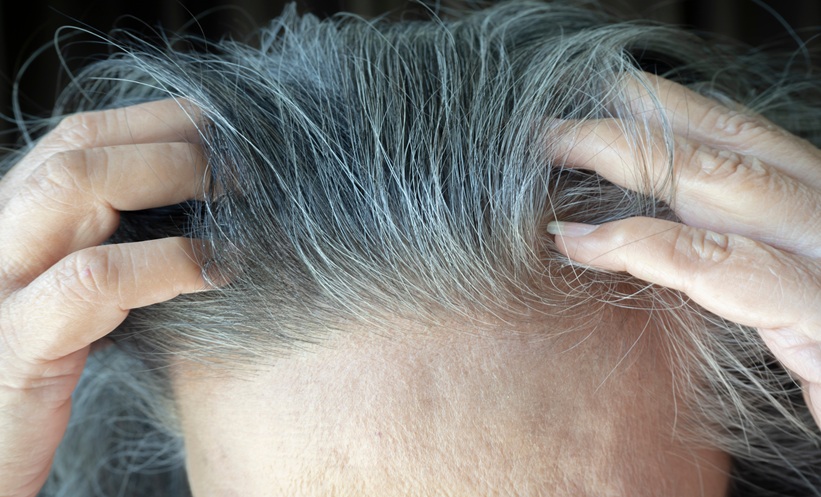
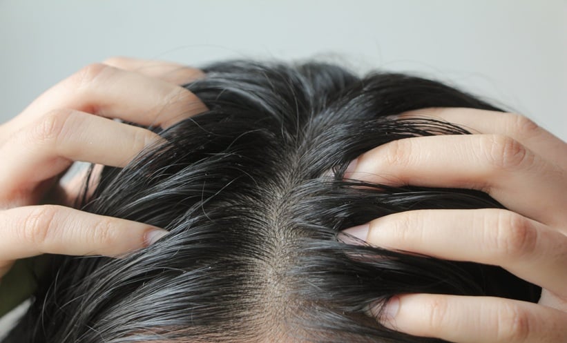
ALOPECIA areata-associated white hair growth (AA-WH) occurred independently of follicular cycling, with ageing, autoimmunity and changes in hair pigmentation identified as associated factors in a large retrospective study. The findings provide further insight into the mechanisms underlying white hair development in patients with alopecia areata (AA), with researchers identifying three potential pathways contributing to the condition.
The study examined clinical characteristics, dermatoscopic findings and histopathological features to better understand the presentation and pathophysiology of AA-WH. Researchers also investigated associations with age, comorbidities and inflammatory markers.
How Common Is White Hair Growth in Alopecia Areata?
AA-WH was identified in 16% of the 5,155 patients included in the study (n=826). Prevalence increased with age and the presence of grey hair, reaching 44.4% among patients aged 55 years and older.
Additional risk factors included older age at AA onset, higher Severity of Alopecia Tool (SALT) scores, coexisting vitiligo, abnormal thyroid function or autoantibodies, and positive antinuclear antibody (ANA) tests.
Clinical presentations included mosaic or uniformly white hair, with localised, diffuse and hairline distribution patterns. Mosaic presentations predominated across AA subtypes, except for alopecia totalis and alopecia universalis.
What Causes White Hair Growth in Alopecia Areata?
Researchers proposed three mechanisms underlying AA-WH: the growth of white vellus hairs, transformation of pigmented terminal hairs, and persistence of pre-existing grey hair. Depigmentation of previously pigmented terminal hairs was also observed.
Compared with patients without AA-WH, affected patients more frequently exhibited hair root depigmentation, trichomalacia, follicular pigment casts, white vellus hair regrowth and weakly pigmented hair bulbs.
Associations with inflammatory and immune markers also varied according to clinical presentation. Diffuse uniform AA-WH was associated with elevated total immunoglobulin E (IgE) and dust-mite-specific IgE in males aged 14 years and younger. In females aged 36 years and older, the same presentation was associated with abnormal thyroid function, thyroid autoantibodies and positive ANA tests.
Hairline or lesion-border AA-WH was associated with elevated total IgE and interleukin-4 levels.
Do Patients Recover from Alopecia Areata-Associated White Hair?
Among patients with follow-up data, 81.7% (353/432) experienced recovery following treatment.
The findings indicate that both depigmentation and repigmentation can occur without evidence of follicular cycling, challenging the assumption that changes in hair pigmentation necessarily depend on the hair growth cycle.
Researchers concluded that AA-WH was associated with ageing, grey hair and autoimmunity across age groups, while allergic features were particularly relevant in younger patients. The identification of distinct pigmentation mechanisms may improve understanding of AA-WH and its clinical presentation.
Reference
Zhang X et al. Alopecia areata-associated white hair growth can occur independent of follicular cycling. Br J Dermatol. 2026;DOI:10.1093/bjd/ljag303.